Dolichosigmoid with Additional Sigmoidal Branches and Displaced Descending Colon
2 Touro College of Osteopahtic Medicine, Harlem, New York, NY 10027, USA
Received: 28-Apr-2021 Accepted Date: May 14, 2021; Published: 24-May-2021, DOI: 10.37532/1308-4038.14(5).126-127
Citation: Kandavalli NB, Wu L, Danquah F, et al. Dolichosigmoid with Additional Sigmoidal Branches and Displaced Descending Colon. Int J Anat Var. 2021;14(5):92-94.
This open-access article is distributed under the terms of the Creative Commons Attribution Non-Commercial License (CC BY-NC) (http://creativecommons.org/licenses/by-nc/4.0/), which permits reuse, distribution and reproduction of the article, provided that the original work is properly cited and the reuse is restricted to noncommercial purposes. For commercial reuse, contact reprints@pulsus.com
Abstract
Sigmoid colon is a part of large intestine and normally measures 15” in length that extends from the pelvicbrim to the third piece of the sacrum where it becomes the rectum. Dolicho sigmoid or redundant sigmoid colon is a term used for sigmoid colon longer than its normal size. The present case had a displaced descending colon and a redundant part of sigmoid colon. During a routine dissection class for medical students, an anatomical variation related to sigmoid colon in an 87-year-old male cadaver was found. The colon was carefully dissected and studied in detail the position, attachment of sigmoidmesocolon, examination of viscera in close proximity and the blood supply to each part of the large intestine. A meticulous review of the literature was conducted as well. An anomalous displacement of descending and sigmoid colons wasfound. The descending colon is normal in length, retroperitoneal and lying close to the midline leaving out a space to its left that was occupied by loops of jejunum and ileum. The sigmoid colon is composed of redundant ascending and normal descending parts. The redundant part of the sigmoid colon is centrally placed as a loop ascending into the peritoneal cavity reaching the splenic flexure at the level of T11 approximately. The descending part of the sigmoid colon crosses the midlineat L1 and runs close to the right midclavicular line continuing as rectum. The total length of sigmoid colon measures 23.2”. The blood supply to the redundant part of the sigmoid comes from a common trunk arising from inferior mesenteric that splits into leftcolic and two additional sigmoid branches. The blood supply to the descending part of sigmoid is supplied by sigmoidal artery coming directly from inferior mesenteric artery. The branching pattern of sigmoidal arteries compared to previous cases in the literature makes this case very unique. Embryology of gut development is complex and often unpredictable, leading to variations in length and position. The growth and rotation of midgut is divided into four stages. An excess of growth of the caudal segment of gut tube in stage 3 can cause redundant sigmoid. This variation could increase the chances of sigmoid volvulus, constipation and may pose risk as the loops may coil around and form a knot leading to obstruction. A redundant loop of sigmoid colon may be asymptomatic or it might lead to urinary, digestive and vascular complications. The descending colon is normal in length but lies close to the midline which may compress the aorta when loaded with feces. Radiologists and surgeons must be well aware of these variations to establish a correct diagnosis and assert the appropriate management when performing colonoscopy, sigmoidoscopy and abdominopelvic procedures.
Introduction
Human large intestine begins at cecum and terminates at anal canal. The colon consists of four segments: ascending, transverse, descending and sigmoid. Normally, the descending colon begins at splenic flexure in the left hypochondriac region, which marks the end of transverse colon. Then it continues vertically and caudally, along the left border of the abdominal cavity and its entire course is located on the left side of jejunum and ileum. The descending colon also forms a narrow longitudinal space with the left abdominal wall called the left paracolic gutter. As the descending colon reaches the pelvic inlet, it becomes the sigmoid colon, which travels medially against the posterior surface of pelvic cavity. As the sigmoid colon extends to S3 vertebral level, it continues inferiorly as the rectum and terminates into the anal canal. The S-shaped sigmoid colon, measures approximately 15’’ in length, is suspended by sigmoid mesocolon and is mobile. The descending and sigmoid colon are supplied by branches of inferior mesenteric artery (IMA): descending by left colic branch and sigmoid by sigmoidal branches [1].
In rare cases, the sigmoid colon is anomalously long and is called redundant sigmoid or dolichosigmoid. Often in these cases, the descending colon is also affected and out of its normal anatomical position. These anomalies are speculated to have significant embryological origins. During the development of human embryos, the intestines go through a complicated herniationrotational process in which errors could occur. This process can be divided into four stages [2,3].
Stage 1: Herniation of primary intestinal loop into the umbilical cord
Stage 2: Rotation of the intestinal loop and elongation of small and large bowels
Stage 3: Retraction of the bowels into the abdominal cavity
Stage 4: Fusion of mesenteries with posterior abdominal wall
Excessive elongation of the large intestine during the second stage may result in abnormally long sigmoid colon. Upon retraction during the third stage, the extra length of the sigmoid colon can be forced into the abdominal cavity and form redundant loops. Additionally, due to faulty rotation in stage two, part of the jejunum and ileum may end up in the paracolic gutter, displacing the descending colon towards the midline. This displacement may become permanent as the mesenteries of descending colon fuse with the posterior abdominal wall.
Medially displaced descending and dolichosigmoid colons could cause many clinical manifestations such as sigmoid volvulus, constipation, urinary and vascular symptoms. Therefore, we are reporting this case to bring significant clinical awareness for surgeons, radiologists and gastroenterologists in their practices [3,4].
Methods and Result Of Dissection
The anomalies were discovered on an 87-year-old male cadaver during a routine anatomical dissecting course for medical students. The cadaver was professionally prepared and the dissection was carefully performed. The ascending and transverse colons, the hepatic flexure and the splenic flexure were normal. The anomalies were found on three structures: the descending colon, the sigmoid colon and the blood supply to the redundant sigmoid colon.
Descending Colon
While the descending colon is retroperitoneal and its length measured 9 inches which is normal, its position was out of the ordinary. In normal cases, as the descending colon takes its course inferiorly towards the pelvic inlet, it runs close to the left lateral abdominal wall, forming a narrow and longitudinal depression in between called the left paracolic gutter. Normally, small intestines and vasculatures are medial to the descending colon. The paracolic gutter usually does not contain any major structure [1].
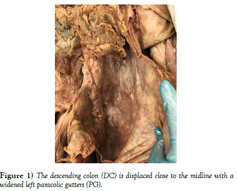
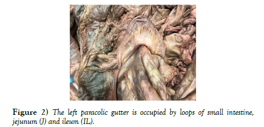
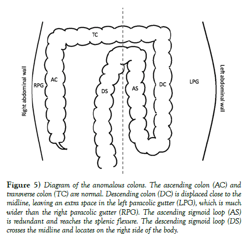
In our case, the entire descending colon was markedly displaced towards the midline, which significantly widened the left paracolic gutter (Figures 1-5). The presence of extra space allowed loops of jejunum and ileum to occupy the enlarged paracolic gutter (Figure 2). Normal colonic configurations, such as haustra, epiploic appendages and tenia coli are present on the descending colon. The circumference also appeared normal. No noticeable pathological change was found in the small bowel as a result of this displacement.
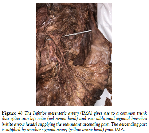
Figure 4) The Inferior mesenteric artery (IMA) gives rise to a common trunk that splits into left colic (red arrow head) and two additional sigmoid branches (white arrow heads) supplying the redundant ascending part. The descending part is supplied by another sigmoid artery (yellow arrow head) from IMA.
Figure 5) Diagram of the anomalous colons. The ascending colon (AC) and transverse colon (TC) are normal. Descending colon (DC) is displaced close to the midline, leaving an extra space in the left paracolic gutter (LPG), which is much wider than the right paracolic gutter (RPG). The ascending sigmoid loop (AS) is redundant and reaches the splenic flexure. The descending sigmoid loop (DS) crosses the midline and locates on the right side of the body.
Sigmoid Colon
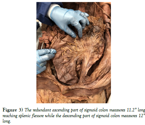
The anomalous sigmoid colon consisted of two segments: a redundant ascending part and a normal descending part. The ascending sigmoidal loop took a sharp turn superiorly as soon as it emerged from the end of the descending colon. It ran continuously upward, parallel but in opposite direction to the descending colon, until it reached the splenic flexure at the vertebral level of T11. The ascending loop was centrally placed and measured 11.2 inches long. Its circumference appeared normal (Figures 3 & 5). The ascending loop of sigmoid colon, or dolichosigmoid, was completely redundant and very rare.
The sigmoid colon then took another sharp turn inferiorly and continued as the descending loop, which looked relatively normal but its position was distorted by the redundant ascending sigmoid. The descending loop was shifted across the midline close to the right midclavicular line as it travelled inferiorly. Upon reaching the vertebral level of L1, it merged with the rectum (Figures 3 & 5). The descending sigmoid was 12 inches long, making the entire sigmoid colon 23.2 inches in length, significantly more than normal values [5].
Blood Supply
Usually, the descending and sigmoid colons are supplied by branches of inferior mesenteric artery: descending colon by the left colic branch and sigmoid colon by sigmoidal branches [1]. In the present case, the redundant ascending part of the sigmoid was perfused by the left colic branch and two sigmoidal branches, all of which originate from a common trunk arising from the inferior mesenteric artery. The descending part is supplied by an additional sigmoidal branch which directly arose from the inferior mesenteric artery (Figure 4).
Discussion
The complex embryological development of the colon makes it susceptible to various anatomical variations in length and position [6]. The sigmoid colon is one of the most variable parts of the large intestines and may cause serious clinical outcomes when anomalously lengthened and displaced [7].
Kanagasuntheram and Loo were pioneers in describing four major classifications for sigmoid colon anomalies. These are I: the presence of an ascending and descending mesocolon, II: the presence of a double hepatic flexure, III: extension of the sigmoid colon into the abdomen, IV: displacement of the sigmoid colon towards the right side [8]. These variations and their associated clinical complications have since been documented. Recently, Zarokosta and colleagues reported an unusual case of lengthening and displacement of the sigmoid colon and mesocolon of a 67-year-old Caucasian male during a hemicolectomy procedure [7]. Additionally, Nayak et al., documented a case of a redundant dolichol-colon in a 65-year-old cadaver during a routine anatomy dissection course [6]. Patel and colleagues have also described a case of sigmoid volvulus in a 14-year-old adolescent girl [9].
In this case study, which holds educational value for clinicians, we have described variations in the length, position, and vascular supply of the descending and sigmoid colon of an 87-year-old male cadaver. The discovery of multiple rare anomalies in the descending and sigmoid colon of this cadaver makes this case study unique [10]. The observation that the descending colon was medially displaced from its normal anatomical position was highly unusual. This resulted in widening of the left paracolic gutter allowing migration of loops of ileum and jejunum into this space. Additionally, the presence of a redundant sigmoid colon, a dolichosigmoid, was also highly peculiar and exceedingly rare [7,9]. Including the redundant segments, the total length of the sigmoid colon was measured at 23.2 inches, which was markedly higher than the averagelength of 6.3cmreported in the literature[5]. Also ofparticular interest was the discovery that the dolichosigmoid colon received its blood supply from variant sigmoidal branches of the inferior mesenteric artery. Since the primordial gut and vasculature are both subject to the error-prone herniation and rotation processes in development, it is logical that anomalies in both structures should occur [11]. This rare finding was observed in this case.
Congenital anomalies in the sigmoid colon have tremendous clinical significance and warrant attention. Due to the sigmoid colon’s close relationships to the pelvic viscera, anomalous course and redundancies risk gastrointestinal, urinary and vascular complications [7]. The most severe of these consequences is a sigmoid volvulus which may lead to obstruction of the descending colon [9]. Furthermore, these anomalies may complicate diagnostic imaging procedures leading to inaccurate interpretation.
Detailed documentation of sigmoid colon anomalies and recognition of their clinical complications is essential for the accurate diagnoses and proper management of these conditions. Therefore, we are presenting this case study to enhance the knowledge of surgeons, gastroenterologists, and radiologists who may encounter these anomalies in their practice.
Declaration of Conflicting Interests
The author(s) declared no potential conflicts of interest with respect to the research, authorship, and/or publication of this article.
Funding
The author(s) did not receive any funding for the research, authorship, and/ or publication of this article.
REFERENCES
- Ibingra CBR. Gross Anatomical variations and congenital anomalies of Surgical importance in hepatobiliary surgery in Uganda. ECAJS. 2006;12:93-8.
- Chiari RS, Shah SA. Sabiston Textbook of Surgery, in Biliary system. St. Louis: WB Saunders. 2008:1474-514.
- Rajguru J, Khare S, Jain S, et al. Variations in the external morphology of Gall Bladder. J Anat Soc Ind. 2012;61:9-12.
- Gore RM, Fulcher AS, Taylor AJ, et al. Textbook of gastrointestinal radiology, in anomalies and anatomic variants of the gallbladder and biliary tract. 2nded. Philadelphia: WB Saunders Co; 2000:1305-20.
- Moore KL, Dalley AF. Clinically oriented anatomy in abdomen. 5thed. Philadelphia: Lippincott Williams & Wilkins; 2006:302.
- Gregor MC, Decker GAG, Du Plessis DJ. Lee MC Gregor’s Synopsis of Surgical Anatomy in the liver & the biliary system. Bombay: KM Verghese& company. 1986:78-103.
- Turner MA, Fulcher AS, Gore RM, et al. Textbook of gastrointestinal radiology in gall bladder and biliary tract: normal anatomy and examination techniques. 2nd ed. Philadelphia: WB Saunders. 2000;2:1250-76.
- Vakili K, Pomfret EA. Biliary Anatomy and embryology. Surg Clin N Am. 2008;88:1159-74.
- Standring S. Gray’s anatomy: The anatomical basis of clinical practice, in gall bladder and biliary tree. 40th ed. Philadelphia: Elsevier Churchill Livingstone; 2008: 69;1177-81.
- Anjankar VP, Panshewdikar PN, Joshi DS, et al. A cadaveric study involving variations in external morphology of gall bladder. Int J Med Res Health Sci. 2013;2:239-42.
- Lichtenstein M, Nicosia AJ. The clinical significance of accessory hepato biliary ducts. Ann Surg. 1955;141:120-4.
- Deutsch AA, Englestein D, Cohen M, et al. Septum of the gall bladder, clinical implications and treatment. Postgrad Med J. 1986;62:453-6.
- Mayo CW, Kendrick DB. Anomalies of the gall bladder: report of a case of left sided floating gall bladder. Arch Surg. 1950;60:668-73.
- Goiney RC, Schoenecer SA, Cyr DA, et al. Sonography of gallbladder duplication and differential considerations. Am J Roentgenol. 1985;145:241-3.
- Barnes S, Nagar H, Levine C, et al. Triple gall bladder: preoperative sonographic diagnosis. J Ultrasound Med. 2004;23:1399-402.
- Bose SM, Sastry RA. Agenesis of gall bladder with choledocho-colonic fistula. Am J Gastroenterol. 1983;78:34-5
- Kabarounders A, Papaziogas B, Atmatzidis K, et al. Hypoplasia of the right hepatic lobe combined with floating gall bladder. Acta Chir Belg. 2003;103:425-7.
- Maeda N, Horie Y, Shiofa G, et al. Hypoplasia of the left lobe associated with floating gall bladder. 1998;45:100-3.


