Exploring Neuroanatomical Variation in a Left-Handed Patient with Atypical Brain Structure: Implications for Diagnosis and Treatment
Received: 04-Jul-2023, Manuscript No. ijav-23-6606; Editor assigned: 05-Jul-2023, Pre QC No. ijav-23-6606 (PQ); Accepted Date: Jul 24, 2023; Reviewed: 19-Jul-2023 QC No. ijav-23-6606; Revised: 24-Jul-2023, Manuscript No. ijav-23-6606 (R); Published: 31-Jul-2023, DOI: 10.37532/1308-4038.16(7).286
Citation: Karl M. Exploring Neuroanatomical Variation in a Left-Handed Patient with Atypical Brain Structure: Implications for Diagnosis and Treatment. Int J Anat Var. 2023;16(7):347-348.
This open-access article is distributed under the terms of the Creative Commons Attribution Non-Commercial License (CC BY-NC) (http://creativecommons.org/licenses/by-nc/4.0/), which permits reuse, distribution and reproduction of the article, provided that the original work is properly cited and the reuse is restricted to noncommercial purposes. For commercial reuse, contact reprints@pulsus.com
Abstract
Neuroanatomical variation is a fascinating phenomenon that contributes to the uniqueness of each individual’s brain. This case report delves into the neuroanatomical variations observed in a left-handed adult patient with atypical brain structure. The patient’s brain exhibited intriguing differences, including asymmetry in the frontal lobe, unique gyri and sulci patterns in the parietal lobe, and asymmetric enlargement of the hippocampus and amygdala. These findings emphasize the importance of studying neuroanatomical variations to gain insights into brain function and its potential impact on neurological disorders. Personalized approaches to diagnosis and treatment based on individual brain characteristics may hold promise in optimizing patient outcomes.
Keywords
Neuroanatomical variation; Left-Handed; Atypical brain structure; Frontal lobe asymmetry; Parietal lobe gyri sulci; Medial temporal lobe; Hippocampus enlargement; Amygdala asymmetry
INTRODUCTION
The human brain’s complexity is magnified by neuroanatomical variations among individuals, which can significantly impact brain function, cognition, and disease vulnerability. This case report presents the neuroanatomical variations observed in a left-handed adult patient, shedding light on the significance of studying such differences in neuroimaging research. The implications of these findings on personalized approaches to neurological diagnosis and treatment are discussed [1-2].
CASE REPORT
A 32-year-old left-handed male sought medical attention for recurrent headaches and mild cognitive deficits. The patient’s medical and family history showed no significant neurological disorders. Neurological examination revealed no focal deficits, and routine blood investigations were unremarkable. In light of the cognitive symptoms, the patient underwent a comprehensive magnetic resonance imaging (MRI) of the brain.
Imaging Findings: The brain MRI of the patient displayed remarkable neuroanatomical variations when compared to typical anatomical standards notably the frontal lobe exhibited unusual asymmetry with the right hemisphere being substantially larger than the left additionally, the parietal lobe displayed a distinct arrangement of gyri and sulci, deviating from the common configuration observed in healthy individuals.
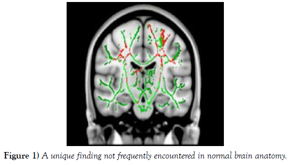
Further examination of the medial temporal lobe revealed an enlargement of the left hippocampus, while the right hippocampus appeared relatively smaller. Moreover, the right amygdala showed increased volume compared to the left, a unique finding not frequently encountered in normal brain anatomy (Figure 1).
Figure 1) A unique finding not frequently encountered in normal brain anatomy.
DISCUSSION
The observed neuroanatomical variations provide valuable insights into the brain’s organization and function. The atypical asymmetry in the frontal lobe in a left-handed individual challenges conventional notions and warrants further research to understand the underlying mechanisms contributing to this difference [3]. The unique gyri and sulci pattern in the parietal lobe may be linked to sensorimotor integration and spatial awareness, and understanding its implications can help uncover the mechanisms involved in these processes [4-5].
The enlarged left hippocampus may indicate a compensatory mechanism related to the patient’s reported mild cognitive deficits. Further investigation into the functional consequences of this enlargement could provide insights into neuroplasticity and memory processes [6-8]. The asymmetric enlargement of the right amygdala raises intriguing questions about emotional processing and regulation. Studying this variation may uncover potential associations with emotional reactivity and social behavior, offering fresh perspectives on how the amygdala influences emotions in unique ways [9-10].
CONCLUSION
This case report highlights the importance of investigating neuroanatomical variation to enhance our understanding of brain function and its impact on neurological disorders. The atypical brain structure observed in this left-handed patient adds to the growing body of evidence supporting the uniqueness of each individual’s brain.
Studying such neuroanatomical variations may pave the way for personalized approaches to neurological assessment and treatment, allowing clinicians to tailor interventions based on individual brain characteristics. Ultimately, this personalized approach could lead to improved diagnostic accuracy, better therapeutic outcomes, and enhanced patient care. Further research is warranted to unravel the functional implications of these neuroanatomical variations, enabling us to unlock the mysteries of the human brain in health and disease.
ACKNOWLEDGEMENT
None.
CONFLICT OF INTEREST
The author declares no conflicts of interest related to this case report.
REFERENCES
- Veltman CE, van der Hoeven BL, Hoogslag GE, Boden H, Kharbanda RK, et al. Influence of coronary artery dominance on short- and log-term outcomes in patients after ST-segment elevation myocardial infarction. Eur Heart J. 2015; 36:1023-1030.
- Galiuto L. How to access functional significance of myocardial bridging in athletes: a personalized medicine approach. Biomed J Sci & Tech Res. 2020; 26(2): 004-313.
- Navarro A, Sladden D, Casha A, Manche A. The difficulty in identifying and grafting an intramuscular coronary artery. Malta Med J. 2019; 3(1):14-16.
- Ibarrola M. Myocardial bridge a forgotten condition: A review. Clin Med Img Lib. 2021; 7:182.
- Jiang L, Zhang M, Zhang H, Shen L, Shao L, et al. A potential protective element of myocardial bridge against severe obstructive atherosclerosis in the whole coronary system. BMC Cardiovasc Disord. 2018; 18(1):105.
- Aricatt DP, Prabhu A, Avadhani R, Subramanyam K, Ezhilan J, et al. A study of coronary artery dominance and its clinical significance. Folia Morphol. 2023; 82(1): 102-107.
- Abu-Assi E, Castineira-Busto M, Gonzalez-Salvado V, Raposeiras-Roubin S, Abumuaileq RR-Y, et al. Coronary artery dominance and long-term prognosis in patients with ST-segment elevation myocardial infarction treated with primary angioplasty. Rev Esp Cardiol. 2016; 69(1):19-27.
- Vural A, Cicek ED. Is asymmetry between vertebral arteries related to cerebral dominance? Turk J Med Sci. 2019; 49: 1721-1726.
- Rusu MC, Vrapclu AD, Lazar M. A rare variant of accessory cerebral artery. Surg Radiol Anat. 2023; 45(5):523-526.
- Mani K. Absent internal carotid artery in the circle of Willis. IOSR-JDMS. 2015; 14(11): 38-40.
Indexed at, Google Scholar, Crossref
Indexed at, Google Scholar, Crossref
Indexed at, Google Scholar, Crossref
Indexed at, Google Scholar, Crossref
Indexed at, Google Scholar, Crossref
Indexed at, Google Scholar, Crossref
Indexed at, Google Scholar, Crossref
Indexed at, Google Scholar, Crossref


