High origin of radial artery – a case report
Enakshi Ghosh1*, Anindya Roy2, Dipankar Kundu3 and Pranab Mukherjee1
1Departments of Anatomy Kolkata, India
2Departments of Physiology Kolkata, India
3R.G. Kar Medical College, Department of Biochemistry, Calcutta Medical College, Kolkata, India
- *Corresponding Author:
- Dr. Enakshi Ghosh
Dept. of Anatomy R.G. Kar Medical College, Kolkata, 700006, India
Tel: +91 890 2494142
E-mail: drenakshighosh@gmail.com
Date of Received: January 30th, 2012
Date of Accepted: July 12th, 2012
Published Online: February 14th, 2013
© Int J Anat Var (IJAV). 2013; 6: 28–30.
[ft_below_content] =>Keywords
high origin, radial artery, variable vascular pattern
Introduction
Brachial artery is the continuation of axillary artery at the lower border of teres major muscle. It gives profunda brachii, nutrient artery to humerus, superior and inferior ulnar collateral arteries and muscular branches in the arm. It ends by dividing into radial and ulnar arteries at the neck of radius.
Radial and ulnar arteries descend in the forearm pursuing a typical course and anastomose with each other in the hand via their branches contributing to the formation of superficial and deep palmar arches [1].
High origin of radial artery is noted in 14.27% of dissected specimens [2] and 9.75% of angiographic examination [3].
Case Report
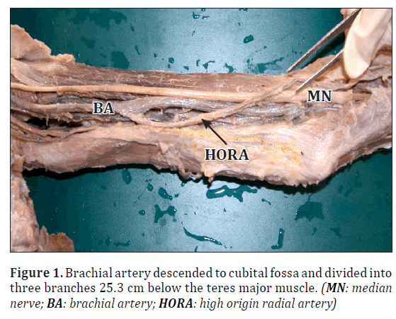
During routine dissection of superior extremity in a 60-year-old male cadaver, high origin of radial artery (HORA) was noted bilaterally. On the left side radial artery originated from the medial side of brachial artery 10.5 cm below the lower border of teres major muscle. The remaining stump of brachial artery descended to cubital fossa and divided in to 3 branches 25.3 cm below the teres major, at the level of the neck of radius. (Figure 1)
Two were muscular branches and the third one was ulnar artery. Further course of ulnar artery was as usual.
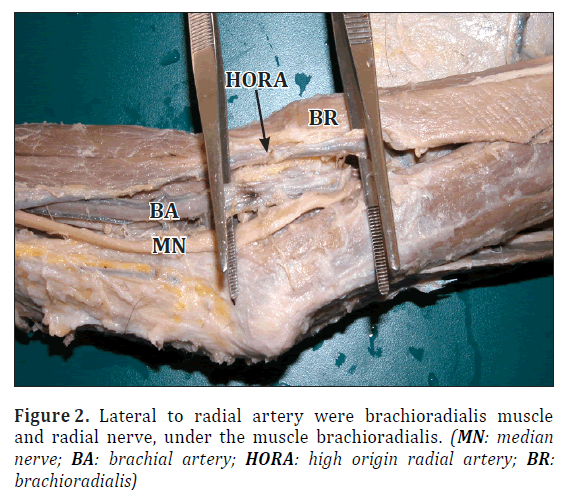
Radial artery crossed brachial artery and median nerve superficially in arm from medial to lateral side and descended to cubital fossa. Radial artery was the fourth structure present in cubital fossa lateral to the tendon of biceps. Still lateral to radial artery were brachioradialis muscle and radial nerve under the brachioradialis muscle. (Figure 2)
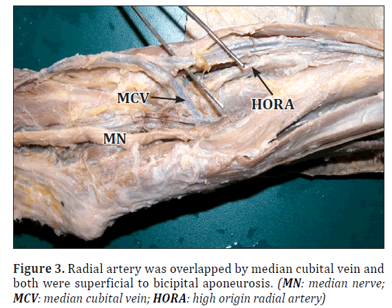
An important finding, worth mentioning here is that median cubital vein continued as one of the venae comitantes of radial artery and was present superficial to radial artery. HORA was overlapped by median cubital vein and both were superficial to bicipital aponeurosis. (Figure 3)
After exit from the apex of cubital fossa radial artery pursued its further course and branching pattern as usual.
Formations of superficial and deep palmar arches were complete; i.e., branches of radial and ulnar arteries anastomosed with each other.
Similar findings were noted on the right side.
Discussion
Embryological explanation
According to Arey variant blood vessels are formed when the developing blood vessels choose an unusual path as developing vessels may fuse with each other forming a single trunk or they develop incompletely. Sometimes a vessel that usually dwindles off may persist and normally persisting vessel may dwindle off. All these changes are dependent on hemodynamics during fetal life [4].
Singer further elaborated the above-mentioned view of Arey and explained five stages for the development of the arteries of the upper limb [5].
Stage 1: Vessels of upper limb develop from subclavian artery that extends all through the length of the limb and ends by supplying digits. Distal part of this vessel is called as anterior interosseous artery.
Stage 2: A median artery arises from subclavian and enlarges to reach and anastomose with anterior interosseous artery; at the same time most distal part of anterior interosseous artery just near the digital branches retrogresses.
Stage 3: Ulnar artery arises as 2nd branch of subclavian artery and enlarges, grows and anastomoses with median artery to form an arch.
Stage 4: Superficial brachial artery arises from original arterial trunk and runs from ulnar to radial side. When artery reaches wrist joint it goes on posterior aspect of wrist and divides over carpus in two branches for dorsum of thumb and index finger.
Stage 5: Four more changes occur: 1) Median artery retrogresses; 2) Anastomotic branch develops just opposite the origin of ulnar artery and connects original trunk to superficial brachial artery; 3) Part of superficial brachial artery above the anastomotic vessel retrogresses; 4) Anastomotic vessel and remaining part of superficial brachial artery becomes radial artery.
According to stage five of Singer, the most likely possibility in present case: The anastomotic branch between the original trunk and the superficial brachial artery did not develop, leading to persistence of embryonic superficial brachial artery. That later on becomes radial artery. Present case shows HORA; i.e., contributed by the proximal part of superficial brachial artery that has persisted and rest of radial artery which pursued its usual course.
HORA forms highest percentage of variations of brachial artery in a study conducted by Karissons, and was reported as 14.27% of dissected material and 9.75% of angiographic examinations [3]. Similarly high incidence of 15% and7.7% has been reported by Anson [6] and also by DdeGaris and Swarttey [7]. Miller in 1939, reported such branch from brachial artery as comperatively lower incidence of 3% only [8]. HORA may be a direct branch of axillary artery as quoted by McCormack et al. [9]. The course of such artery in the arm is lying lateral to median nerve but as it progresses in the forearm it has an usual course, that has been reported by Karlsson and Niechajev [3], Anson [6] and Huber [10]. Similar findings are also supported by McCormack et al. [9], and also by Compta[11]. Karlsson & Niechajev [3] noted incidence of HORA in dissected specimens as high as 10% and mostly similar incidence of 9.7% in angiographic examinations. In the present case at its origin the HORA crosses the brachial artery and median nerve from medial to lateral side, and is placed superficial to these structures. Hence it can be mistaken as a muscular branch during open reduction of fracture middle 1/3rd of humerus. It may be clamped or ligated leading to compromised vascular supply particularly to the thumb which may derive its sole supply from branch of radial artery in 18.46% of cases [12]. Here we get presence of complete palmar arch, i.e., branches of radial and ulnar arteries anastomosed with each other at superficial and deep palmar arch [13]. HORA is an additional content of cubital fossa, placed deep to median cubital vein. While cannulating median cubital vein for blood sampling either for donation or routine diagnostic purpose or intravenous injection; the slightly deeply placed needle may enter HORA and lead to serious complications like hemorrhage, arteriovenous fistula or an intravenous drug may be wrongly administered intraarterially due to their close proximity.
Radial artery is harvested as a graft for coronary artery bypass surgery. HORA coupled with incomplete palmar arches can cause compromised supply to the thumb leading to its gangrene or amputation as rare postoperative complication.
Position of angiographic catheter for study of vascular pattern in upper extremity can be done perfectly only with the knowledge of presence of HORA. A catheter placed near the terminal end of brachial artery for study of vascular pattern in the forearm may miss HORA, finally leading to wrong interpretation of angiographic findings as far as radial artery is concerned.
Knowledge of variations in vascular pattern of upper extremity is crucial and necessary to avoid complications encountered during venotomy, surgical procedures and ambiguity during radiological and angiographic diagnostic procedures.
The study of vascular pattern of upper extremity which includes all the arteries and their branches from axillary artery to digital branches of radial and ulnar that supply fingers should be undertaken to give complete justification and clinical relevance to the above mentioned findings.
References
- Standring S, ed. Gray’s Anatomy. 40th Ed., Edinburgh, Churchill Livingstone. 2008; 852.
- Gessini L, Jandolo B, Pietrangeli A. Entrapment neuropathies of the median nerve at and above the elbow. Surg Neurol. 1983; 19: 112–116.
- Karlsson S, Niechajev IA. Arterial anatomy of the upper extremity. Acta Radiol Diagn (Stockh). 1982; 23: 115–121.
- Aray L. Developmental Anatomy. 6th Ed., Philadelphia, W. B. Saunders Co. 1953: 375–377.
- Singer E. Embryological pattern persisting in the arteries of the arm. Anat Rec. 1933; 55: 406–413.
- Anson BJ. Morris’ Human Anatomy. New York, McGraw Hill Book C. 1966; 708–724.
- De Garis CF, Swarttey WB. The axillary artery in white and negro stocks. Am J Anat. 1963; 41: 353–397.
- Miller RA. Observations upon the arrangement of axillary artery and brachial plexus. Am J Anat. 1939; 64: 143–163.
- McCormack LJ, Cauldwell EW, Anson BJ. Brachial and antebrachial arterial patterns; a study of 750 extremities. Surg Gynecol Obstet. 1953; 96: 43–54.
- Huber GC. Piersol’s Human Anatomy. 9th Ed., Philadelphia, J. B. Lippincott Co. 1930; 767–791.
- Gonzalez-Compta X. Origin of the radial artery from the axillary artery and associated hand vascular anomalies. J Hand Surg Am. 1991; 16: 293–296.
- Chimmalgi M, Choudnary R, Tuli A, Chhiber SR, Humbarwadi RS. Arterial supply of thumb. Anatomica Karnataka. 2002; 23: 23–25.
- Coleman SS, Anson BJ. Arterial patterns in the hand based upon a study of 650 specimens. Surg Gynecol Obstet. 1961; 113: 409–424.
Enakshi Ghosh1*, Anindya Roy2, Dipankar Kundu3 and Pranab Mukherjee1
1Departments of Anatomy Kolkata, India
2Departments of Physiology Kolkata, India
3R.G. Kar Medical College, Department of Biochemistry, Calcutta Medical College, Kolkata, India
- *Corresponding Author:
- Dr. Enakshi Ghosh
Dept. of Anatomy R.G. Kar Medical College, Kolkata, 700006, India
Tel: +91 890 2494142
E-mail: drenakshighosh@gmail.com
Date of Received: January 30th, 2012
Date of Accepted: July 12th, 2012
Published Online: February 14th, 2013
© Int J Anat Var (IJAV). 2013; 6: 28–30.
Abstract
High origin of radial artery was seen bilaterally in a 60-year-old male cadaver during routine dissection in the Department of Anatomy, R.G. Kar Medical College, Kolkata, India. Knowledge of variations in vascular pattern is clinically important because of routinely performed angiographic procedures and surgical treatment. High origin of radial artery is noted in 14.27% of dissected specimens and 9.75% of angiographic examination.
-Keywords
high origin, radial artery, variable vascular pattern
Introduction
Brachial artery is the continuation of axillary artery at the lower border of teres major muscle. It gives profunda brachii, nutrient artery to humerus, superior and inferior ulnar collateral arteries and muscular branches in the arm. It ends by dividing into radial and ulnar arteries at the neck of radius.
Radial and ulnar arteries descend in the forearm pursuing a typical course and anastomose with each other in the hand via their branches contributing to the formation of superficial and deep palmar arches [1].
High origin of radial artery is noted in 14.27% of dissected specimens [2] and 9.75% of angiographic examination [3].
Case Report
During routine dissection of superior extremity in a 60-year-old male cadaver, high origin of radial artery (HORA) was noted bilaterally. On the left side radial artery originated from the medial side of brachial artery 10.5 cm below the lower border of teres major muscle. The remaining stump of brachial artery descended to cubital fossa and divided in to 3 branches 25.3 cm below the teres major, at the level of the neck of radius. (Figure 1)
Two were muscular branches and the third one was ulnar artery. Further course of ulnar artery was as usual.
Radial artery crossed brachial artery and median nerve superficially in arm from medial to lateral side and descended to cubital fossa. Radial artery was the fourth structure present in cubital fossa lateral to the tendon of biceps. Still lateral to radial artery were brachioradialis muscle and radial nerve under the brachioradialis muscle. (Figure 2)
An important finding, worth mentioning here is that median cubital vein continued as one of the venae comitantes of radial artery and was present superficial to radial artery. HORA was overlapped by median cubital vein and both were superficial to bicipital aponeurosis. (Figure 3)
After exit from the apex of cubital fossa radial artery pursued its further course and branching pattern as usual.
Formations of superficial and deep palmar arches were complete; i.e., branches of radial and ulnar arteries anastomosed with each other.
Similar findings were noted on the right side.
Discussion
Embryological explanation
According to Arey variant blood vessels are formed when the developing blood vessels choose an unusual path as developing vessels may fuse with each other forming a single trunk or they develop incompletely. Sometimes a vessel that usually dwindles off may persist and normally persisting vessel may dwindle off. All these changes are dependent on hemodynamics during fetal life [4].
Singer further elaborated the above-mentioned view of Arey and explained five stages for the development of the arteries of the upper limb [5].
Stage 1: Vessels of upper limb develop from subclavian artery that extends all through the length of the limb and ends by supplying digits. Distal part of this vessel is called as anterior interosseous artery.
Stage 2: A median artery arises from subclavian and enlarges to reach and anastomose with anterior interosseous artery; at the same time most distal part of anterior interosseous artery just near the digital branches retrogresses.
Stage 3: Ulnar artery arises as 2nd branch of subclavian artery and enlarges, grows and anastomoses with median artery to form an arch.
Stage 4: Superficial brachial artery arises from original arterial trunk and runs from ulnar to radial side. When artery reaches wrist joint it goes on posterior aspect of wrist and divides over carpus in two branches for dorsum of thumb and index finger.
Stage 5: Four more changes occur: 1) Median artery retrogresses; 2) Anastomotic branch develops just opposite the origin of ulnar artery and connects original trunk to superficial brachial artery; 3) Part of superficial brachial artery above the anastomotic vessel retrogresses; 4) Anastomotic vessel and remaining part of superficial brachial artery becomes radial artery.
According to stage five of Singer, the most likely possibility in present case: The anastomotic branch between the original trunk and the superficial brachial artery did not develop, leading to persistence of embryonic superficial brachial artery. That later on becomes radial artery. Present case shows HORA; i.e., contributed by the proximal part of superficial brachial artery that has persisted and rest of radial artery which pursued its usual course.
HORA forms highest percentage of variations of brachial artery in a study conducted by Karissons, and was reported as 14.27% of dissected material and 9.75% of angiographic examinations [3]. Similarly high incidence of 15% and7.7% has been reported by Anson [6] and also by DdeGaris and Swarttey [7]. Miller in 1939, reported such branch from brachial artery as comperatively lower incidence of 3% only [8]. HORA may be a direct branch of axillary artery as quoted by McCormack et al. [9]. The course of such artery in the arm is lying lateral to median nerve but as it progresses in the forearm it has an usual course, that has been reported by Karlsson and Niechajev [3], Anson [6] and Huber [10]. Similar findings are also supported by McCormack et al. [9], and also by Compta[11]. Karlsson & Niechajev [3] noted incidence of HORA in dissected specimens as high as 10% and mostly similar incidence of 9.7% in angiographic examinations. In the present case at its origin the HORA crosses the brachial artery and median nerve from medial to lateral side, and is placed superficial to these structures. Hence it can be mistaken as a muscular branch during open reduction of fracture middle 1/3rd of humerus. It may be clamped or ligated leading to compromised vascular supply particularly to the thumb which may derive its sole supply from branch of radial artery in 18.46% of cases [12]. Here we get presence of complete palmar arch, i.e., branches of radial and ulnar arteries anastomosed with each other at superficial and deep palmar arch [13]. HORA is an additional content of cubital fossa, placed deep to median cubital vein. While cannulating median cubital vein for blood sampling either for donation or routine diagnostic purpose or intravenous injection; the slightly deeply placed needle may enter HORA and lead to serious complications like hemorrhage, arteriovenous fistula or an intravenous drug may be wrongly administered intraarterially due to their close proximity.
Radial artery is harvested as a graft for coronary artery bypass surgery. HORA coupled with incomplete palmar arches can cause compromised supply to the thumb leading to its gangrene or amputation as rare postoperative complication.
Position of angiographic catheter for study of vascular pattern in upper extremity can be done perfectly only with the knowledge of presence of HORA. A catheter placed near the terminal end of brachial artery for study of vascular pattern in the forearm may miss HORA, finally leading to wrong interpretation of angiographic findings as far as radial artery is concerned.
Knowledge of variations in vascular pattern of upper extremity is crucial and necessary to avoid complications encountered during venotomy, surgical procedures and ambiguity during radiological and angiographic diagnostic procedures.
The study of vascular pattern of upper extremity which includes all the arteries and their branches from axillary artery to digital branches of radial and ulnar that supply fingers should be undertaken to give complete justification and clinical relevance to the above mentioned findings.
References
- Standring S, ed. Gray’s Anatomy. 40th Ed., Edinburgh, Churchill Livingstone. 2008; 852.
- Gessini L, Jandolo B, Pietrangeli A. Entrapment neuropathies of the median nerve at and above the elbow. Surg Neurol. 1983; 19: 112–116.
- Karlsson S, Niechajev IA. Arterial anatomy of the upper extremity. Acta Radiol Diagn (Stockh). 1982; 23: 115–121.
- Aray L. Developmental Anatomy. 6th Ed., Philadelphia, W. B. Saunders Co. 1953: 375–377.
- Singer E. Embryological pattern persisting in the arteries of the arm. Anat Rec. 1933; 55: 406–413.
- Anson BJ. Morris’ Human Anatomy. New York, McGraw Hill Book C. 1966; 708–724.
- De Garis CF, Swarttey WB. The axillary artery in white and negro stocks. Am J Anat. 1963; 41: 353–397.
- Miller RA. Observations upon the arrangement of axillary artery and brachial plexus. Am J Anat. 1939; 64: 143–163.
- McCormack LJ, Cauldwell EW, Anson BJ. Brachial and antebrachial arterial patterns; a study of 750 extremities. Surg Gynecol Obstet. 1953; 96: 43–54.
- Huber GC. Piersol’s Human Anatomy. 9th Ed., Philadelphia, J. B. Lippincott Co. 1930; 767–791.
- Gonzalez-Compta X. Origin of the radial artery from the axillary artery and associated hand vascular anomalies. J Hand Surg Am. 1991; 16: 293–296.
- Chimmalgi M, Choudnary R, Tuli A, Chhiber SR, Humbarwadi RS. Arterial supply of thumb. Anatomica Karnataka. 2002; 23: 23–25.
- Coleman SS, Anson BJ. Arterial patterns in the hand based upon a study of 650 specimens. Surg Gynecol Obstet. 1961; 113: 409–424.


