Is The Flexor Digiti Minimi Brevis Muscle of the Foot Belong To First Layer or Third Layer
Received: 24-Jun-2021 Accepted Date: Jul 12, 2021; Published: 22-Jul-2021, DOI: 10.37532/1308-4038.14(7).95-96
Citation: Karip B Köse OO. Is The Flexor Digiti Minimi Brevis Muscle of the Foot Belong To First Layer or Third Layer? Int J Anat Var. 2021;14(7):110-110.
This open-access article is distributed under the terms of the Creative Commons Attribution Non-Commercial License (CC BY-NC) (http://creativecommons.org/licenses/by-nc/4.0/), which permits reuse, distribution and reproduction of the article, provided that the original work is properly cited and the reuse is restricted to noncommercial purposes. For commercial reuse, contact reprints@pulsus.com
Abstract
The plantar area of the foot is known as very complicated in terms of muscle layers. The first layer’s muscle, abductor digiti minimi and the third layer’s muscle, flexor digiti minimi brevis are so familiar and located closer to eachother. According to anatomical aspect; abductor digiti minimi muscle lies on the flexor digitimi minimi brevis but in the case that we work on, flexor digiti minimi brevis muscle lies on the abductor digiti minimi. The reasons may followed as; the function of the lateral plantar side, genetic and environmental conditions as well.
Keywords
Abductor digiti minimi muscle; Flexor digiti minimi brevis muscle; Foot
Introduction
The Defining the plantar side of the foot muscles can be difficult for the dissectors, students or someones who try to learn anatomy. That is why the plantar muscles subdivided into 4 layers and all these layers evaluated in their nature. Plantar aspect of the foot contains so many branches of arteries, nerves and veins and one has to be careful about the applications for this area. The muscles of this area may have the variations so the structures told as variants are important for the clinical operations for the surgeons especially. In the lateral plantar side of the foot consists of the abductor and flexor muscles. As specified; ther abducts and flexes the toe. Abductor digiti minimi muscle is originated from the calcaneal tuberosity and intermuscular septum and is inserted into the base of fifth metatarsal bone. It has the important relation with the flexor digiti minimi brevis muscle that is located under. Flexor digiti minimi brevis muscle is originated from the base of fifth mettarsal bone and long plantar ligament and is inserted into fifth digit’ s proximal phalanx. During the surgical prosedure, cilinicians generally state that one of the most easily accessible muscles in foot is abductor digiti minimi and that is why this muscle is often used as flap in the surgery [1]. Abductor digimi minimi muscle is also used as flap for lateral plantar side of the foot in diabetic patients because of the huge vounds [2]. There also can be seen the repetitive traumas on the lateral malleolar side of the foot because of the broken bones. If the abductor digiti minimi is suitable for use, it can be a great flap for all these traumatic injuries [3]. Flexor digiti minimi brevis muscle of the foot is located at the lateral plantar side and also one of the most variable muscles. Between its variations the most popular one is that there are lateral and medial heads found together [4].
Case Report
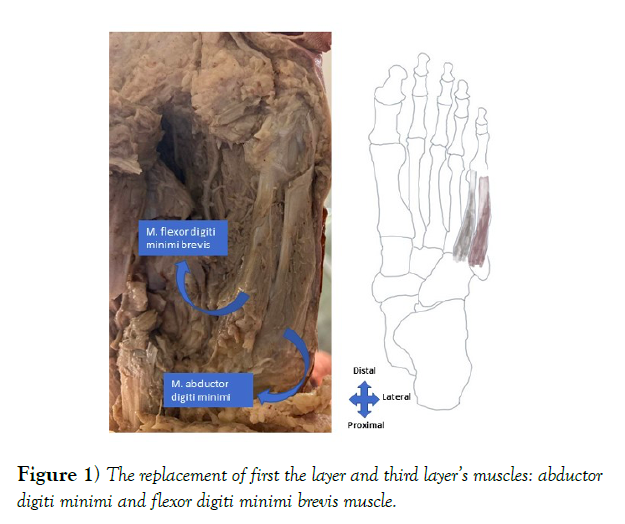
During the routin cadaver dissection, we observed that abductor digiti minimi and flexor digiti minimi brevis muscles were not in their normal positions such that there was a variable order between them. As shown in Figure 1, the abductor digiti minimi muscle lies down under the flexor digiti minimi brevis. The origin of the flexor digitimi brevis muscle was the base of fifth metatarsal bone and was inserted into the base of fifth proximal phalanx. It was lying down the medial and upper side of the abductor digiti minimi. The abductor digiti minimi muscle was originated from the calcaneal tuberosity of the foot and it was also inserted into base of fifth metatarsal and the proximal phalanx of the fifth digit.
Discussion and Conclusion
In terms of clinical importance of the muscles; the normal and variable forms have to know better. Some muscles in human body especially can be used as flap and for the foot the abductor digiti minimi and flexor digiti minimi brevis muscles are so favourite in some diseases. Normally the first layer muscle is located at the upper part of the flexor digiti minimi brevis but we observed that there was a reverse condition. The reason may be genetic or the state of use. We conclude that in clinical conditions abductor digiti minimi and flexor digiti minimi brevis muscles can not be confused. These two can be found as we declared. The study can be improved with by using many numbers of cadavers.
REFERENCES
- Eckardt A, Himmelhan ZR, Oper H, et al. Redaktion Amputationen am Rückfuß Operative Techniken. Orthop Traumatol. 2011;23:265-279
- Ramanujam CL, Suto AC, Zgonis T. Modification of the abductor digiti minimi muscle flap for soft tissue coverage of the diabetic foot. Journal of Wound Care. 2020;29:32-36.
- Elfeki B, Eun S. Lateral malleolar defect coverage using abductor digiti minimi muscle flap. Annals of Plastic Surgery. 2019;83:50-54.
- Mehta V, Gupta V, Arora J, et al. An atypical composition of adductor hallucis co-existent with an accessory plantar muscle and duplication of flexor digiti minimi pedis. Clinica Terapeutica. 2011;162:361-363.


