Preperitoneal pelvic kidney: Revisiting the significance of variant anatomy to the clinician of the future
Afadhali Denis Russa*
Department of Anatomy, School of Medicine, Muhimbili University of Health Sciences and Allied Sciences, Tanzania
- *Corresponding Author:
- Dr. Afadhali Denis Russa
Department of Anatomy
School of Medicine
Muhimbili University of Health Sciences and Allied Sciences, Tanzania
Telephone +255 755 524 771
E-mail: drussa@muhas.ac.tz
Published Online: 2 June 2017
Citation: Russa AD. Preperitoneal pelvic kidney: Revisiting the significance of variant anatomy to the clinician of the future. Int J Anat Var. 2017;10(2):021-23.
© This open-access article is distributed under the terms of the Creative Commons Attribution Non-Commercial License (CC BY-NC) (http:// creativecommons.org/licenses/by-nc/4.0/), which permits reuse, distribution and reproduction of the article, provided that the original work is properly cited and the reuse is restricted to noncommercial purposes. For commercial reuse, contact reprints@pulsus.com
[ft_below_content] =>Keywords
Pelvic, Kidney, Variant, Anatomy, Significance
Introduction
The kidneys are retroperitoneal organs applied to the posterior abdominal wall on either side of the vertebral bodies from upper border of T12 to L3. Grossly, the kidneys are bean-shaped structures and weigh about 150 g in the male and about 135 g in the female. Kidneys measure 10-12 cm in length, 5-7 cm in width, and 2-3 cm in thickness. While the normal kidneys show slight variations in position in their retroperitoneal abdominal location, ectopic kidneys are a common encounter in autopsies and the clinical set up. It is estimated that up to 40% of the urinary pathologies are a result of these anatomical variations [1]. Renal ectopia have been associated with different pathologies ranging from incompatibility with life, increased incidences of renal cancers, systemic conditions such are hypertension to asymptomatic normal functioning kidneys [2,3]. Despite these functional and clinical significances, these variations are scarcely emphasized in the anatomy books taught to the medical students—the surgeons of the future. Additionally, there are few clinical guidelines and management protocols that take variant renal anatomy into consideration. In the present paper we are reporting a rare form of renal ectopia presenting with pre-peritoneal location, multiple structural variations and variant vasculature. We also discuss possible functional implications and surgical challenges arising from renal ectopia.
Case Report
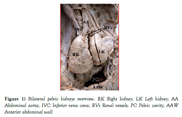
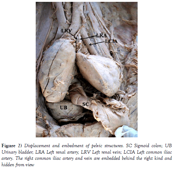
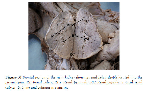
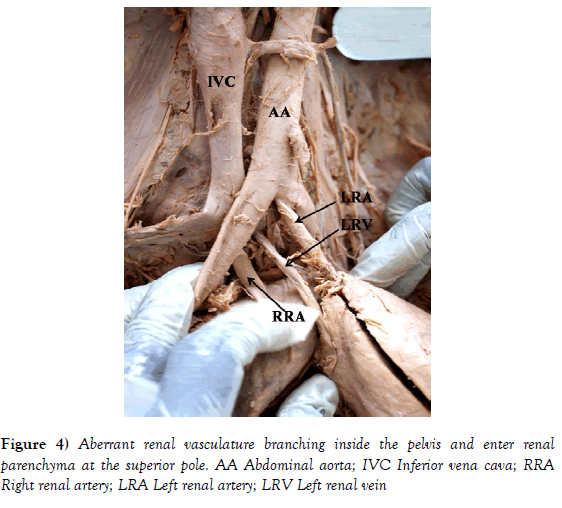
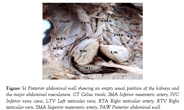
During the dissection sessions of the medical students at the Muhimbili University of Health and Allied Sciences (MUHAS) we observed a case of bilateral pelvic kidneys with aberrant size, structure and vascular supply in a male cadaver aged 53 years whose death was otherwise not of a renal cause. The kidneys were relatively larger (12 × 6 × 4 cm) with the right kidney longer and slender while the left was triangular in shape. Both kidneys occupied the pelvic cavity and were pre-peritoneal. The long axis of the right kidney lay along the right pelvic inlet. The superior poles of both kidneys pressed against each other leading to atypical morphology (Figure 1). The two kidneys being pelvic in position, lost their retroperitoneal position and they overhung the sigmoid colon and the urinary bladder and, in so doing, displacing them towards the floor of the pelvis. The kidney also pressed the common iliac vessels posteriorly, against the borders of the pelvic inlet (Figure 2). Internally the renal structure was unusual with the renal pelvis deep into the parenchyma and presenting with atypical renal pyramids and pelvis (Figure 3). The renal vasculature was also aberrant in origin, course and termination. The left renal artery took origin from the bifurcation of the abdominal aorta into common iliac and measured 5.1 cm whereas the right renal artery stemmed from the right common iliac artery and measured 4.0 cm. The iliac veins took origin from inferior vena cava bifurcation into common iliac veins. Both the arteries and veins entered the renal tissue at the superior pole as the kidneys did not possess conventional hila. The arteries had an unusual vertical course as they descended to supply the pelvic kidneys. Renal veins were not measured but from the photographs, their length and course followed those of the arteries. The testicular vessels also showed variations on origin. The renal arteries took origin from respective iliac vessels. The right testicular vein originated as usual as an anterior branch of the inferior vena cove. The left testicular vein—which normally stem from left renal vein—originated together with the left suprarenal vein as a common stem from the inferior vena cover in the abdomen. The course of testicular veins was unaffected. Ureters were short with their entire length located in the pelvis and laterally displaced by the pelvis kidneys. Other abdominal vasculatures were also less affected including the celiac trunk and superior and inferior mesenteric arteries (Figures 4 and 5) [1-7].
Figure 5: Posterior abdominal wall showing an empty usual position of the kidneys and the major abdominal vasculature. CT Celiac trunk; IMA Inferior mesenteric artery; IVC Inferior vena cava; LTV Left testicular vein; RTA Right testicular artery; RTV Right testicular vein; SMA Superior mesenteric artery; PAW Posterior abdominal wall
On a lower level, at the same height as the aorta bifurcation arise another anomalous polar artery with 4.93 mm in size and 75.29 mm in length from its origin until its entrance in the renal hilum following a rectilinear trajectory until the inferior polo of the right kidney (Figure 2).
Discussion
Variant renal anatomy is extremely common and in many cases poses surgical and imaging challenges [1,5]. The present case is a prototype of such cases. Similar findings have been reported previously. Renal pelvic ectopia occurs as a result of a defective or an incomplete ascent of the developing kidneys [3]. Such defective ascent might explain the unusual bigger size of the kidneys in the present case. Clinically the unusually massive and irregular kidneys pressing the iliac vessels against the bony pelvis could easily cause vascular obstructions leading to several pathologies of blood circulations. Unlike the much spacious abdominal cavity, the pelvic cavity has a limited space and the occurrence of the kidneys in such aberrant location may lead to displacement and embedment of the pelvic viscera. Displacement of the ureters by the pelvic kidneys may lead to dysuria and other urological pathologies [8]. Chances of renal vessel obstruction are high due to the abnormal course of the renal vessels as they cross the pelvic inlet. Pelvic nerve entrapment is also a likely feature. Generally the inborn urinary conditions occur in 1:25 of all live births [4] and accounts to about 40% of all urinary system pathologies [8]. Of these, cases of renal ectopia are estimated at 1:500 to 1:110 [1]. Pelvic type alone accounts to 1:2500 of all live births [6]. These major patterns and occurrence of variant renal anatomy need to be well documented and included in the anatomy test books for medical and specializing residents in different clinical specialties. For instance, these anatomical variations are not mentioned in three of the common standard anatomy text books for medical students and surgical residents worldwide—Gray’s Anatomy for Students, Moore’s Clinically Oriented Anatomy and Snell’s Clinical Anatomy by Regions. While the more extensive Gray’s Anatomy puts some appreciable level of emphasis on the variant renal anatomy [6], the book is less used as a standard text book for anatomy teaching. Silence towards such potential abnormal findings creates a knowledge gap and may depict a distorted understanding of the functional anatomy by the future surgeon and hence setting a shaky foundation in managing cases of the genitourinary system.
Conclusion
Reports of extreme renal variations in structure and location are of radiological and surgical significances. Urologists and nephrologists and other clinicians need to have in mind these relatively common variations of the kidney for successful plans and outcomes of the clinical procedures. Due to its common occurrence, renal ectopia should be emphasized in normal anatomy class for medical students and surgical residents. Routine clinical and surgical guidelines should also take these relatively common variations into considerations
References
- Belsare SM, Chimmalgi M, Vaidya SA, et al. Ectopic Kidney and associated anomalies: A Case Report J Anat. Soc. 2002;51:236-8.
- Gülsün M, Balkanci F, Cekirge S, et al. Pelvic kidney with an unusual blood supply: angiographic findings. Surg Radiol Anat. 2000;22:59-61.
- Hegazy AA. Clinical Embryology for Medical Students and Postgraduate Doctors. Berlin: Lap Lambert Academic Publishing, 2014.
- Moore KL, Persaud TVN. The Developing Human, Clinically Oriented Embryology. 8th Ed., Philadelphia, Saunders. 2008;244-60.
- Raman SS, Pojchamarnwiputh S, Muangsomboon K, et al. Surgically Relevant Normal and Variant Renal Parenchymal and Vascular Anatomy in Preoperative 16-MDCT Evaluation of Potential Laparoscopic Renal Donors. Am J Roentgenol. 2007;188:105-14.
- Standring S, Jeremiah HC, Borley NR, et al. eds. Gray’s Anatomy. The Anatomical Basis of Clinical Practice. 39th Ed., Edinburgh, London, New York, Oxford, Philadelphia, St. Louis, Sydney, Toronto, Elsevier Churchill Livingstone. 2005;1269–86.
- Wein AJ, Kavoussi LR, Novick AC, et al. Campbell-Walsh Urology. 9th Edn., Saunders. 2007;I:24.
- Williams PL, Bannister LH, Warwick R, et al. Embryology & Development, Urinary System in “Gray’s anatomy,” 38th Ed, Churchill Livingstone, London, UK. 1995;199-204.
Afadhali Denis Russa*
Department of Anatomy, School of Medicine, Muhimbili University of Health Sciences and Allied Sciences, Tanzania
- *Corresponding Author:
- Dr. Afadhali Denis Russa
Department of Anatomy
School of Medicine
Muhimbili University of Health Sciences and Allied Sciences, Tanzania
Telephone +255 755 524 771
E-mail: drussa@muhas.ac.tz
Published Online: 2 June 2017
Citation: Russa AD. Preperitoneal pelvic kidney: Revisiting the significance of variant anatomy to the clinician of the future. Int J Anat Var. 2017;10(2):021-23.
© This open-access article is distributed under the terms of the Creative Commons Attribution Non-Commercial License (CC BY-NC) (http:// creativecommons.org/licenses/by-nc/4.0/), which permits reuse, distribution and reproduction of the article, provided that the original work is properly cited and the reuse is restricted to noncommercial purposes. For commercial reuse, contact reprints@pulsus.com
Abstract
The kidneys are retroperitoneal organs applied to the posterior abdominal wall. Renal ectopia and structural alterations are of clinical significance. Most renal ectopia occurs in the pelvis and has a strong association with genitourinary pathologies. I the present study we report a rare case of bilateral pre-peritoneal pelvic renal ectopia with aberrant size, structure and renal vasculature. The ectopic kidneys displaced the pelvic viscera inferiorly. This pre-peritoneal position of the kidney is rarely reported. The aberrant kidneys bore an atypical internal structure with renal pelvis deeply situated into the parenchyma. These anatomical variations are not mentioned in the common standard anatomy text books for medical students and surgical residents. Due to their common occurrence, however, renal ectopia should be emphasized in normal anatomy class for medical students and surgical residents. Routine clinical guidelines should take into considerations these variations.
-Keywords
Pelvic, Kidney, Variant, Anatomy, Significance
Introduction
The kidneys are retroperitoneal organs applied to the posterior abdominal wall on either side of the vertebral bodies from upper border of T12 to L3. Grossly, the kidneys are bean-shaped structures and weigh about 150 g in the male and about 135 g in the female. Kidneys measure 10-12 cm in length, 5-7 cm in width, and 2-3 cm in thickness. While the normal kidneys show slight variations in position in their retroperitoneal abdominal location, ectopic kidneys are a common encounter in autopsies and the clinical set up. It is estimated that up to 40% of the urinary pathologies are a result of these anatomical variations [1]. Renal ectopia have been associated with different pathologies ranging from incompatibility with life, increased incidences of renal cancers, systemic conditions such are hypertension to asymptomatic normal functioning kidneys [2,3]. Despite these functional and clinical significances, these variations are scarcely emphasized in the anatomy books taught to the medical students—the surgeons of the future. Additionally, there are few clinical guidelines and management protocols that take variant renal anatomy into consideration. In the present paper we are reporting a rare form of renal ectopia presenting with pre-peritoneal location, multiple structural variations and variant vasculature. We also discuss possible functional implications and surgical challenges arising from renal ectopia.
Case Report
During the dissection sessions of the medical students at the Muhimbili University of Health and Allied Sciences (MUHAS) we observed a case of bilateral pelvic kidneys with aberrant size, structure and vascular supply in a male cadaver aged 53 years whose death was otherwise not of a renal cause. The kidneys were relatively larger (12 × 6 × 4 cm) with the right kidney longer and slender while the left was triangular in shape. Both kidneys occupied the pelvic cavity and were pre-peritoneal. The long axis of the right kidney lay along the right pelvic inlet. The superior poles of both kidneys pressed against each other leading to atypical morphology (Figure 1). The two kidneys being pelvic in position, lost their retroperitoneal position and they overhung the sigmoid colon and the urinary bladder and, in so doing, displacing them towards the floor of the pelvis. The kidney also pressed the common iliac vessels posteriorly, against the borders of the pelvic inlet (Figure 2). Internally the renal structure was unusual with the renal pelvis deep into the parenchyma and presenting with atypical renal pyramids and pelvis (Figure 3). The renal vasculature was also aberrant in origin, course and termination. The left renal artery took origin from the bifurcation of the abdominal aorta into common iliac and measured 5.1 cm whereas the right renal artery stemmed from the right common iliac artery and measured 4.0 cm. The iliac veins took origin from inferior vena cava bifurcation into common iliac veins. Both the arteries and veins entered the renal tissue at the superior pole as the kidneys did not possess conventional hila. The arteries had an unusual vertical course as they descended to supply the pelvic kidneys. Renal veins were not measured but from the photographs, their length and course followed those of the arteries. The testicular vessels also showed variations on origin. The renal arteries took origin from respective iliac vessels. The right testicular vein originated as usual as an anterior branch of the inferior vena cove. The left testicular vein—which normally stem from left renal vein—originated together with the left suprarenal vein as a common stem from the inferior vena cover in the abdomen. The course of testicular veins was unaffected. Ureters were short with their entire length located in the pelvis and laterally displaced by the pelvis kidneys. Other abdominal vasculatures were also less affected including the celiac trunk and superior and inferior mesenteric arteries (Figures 4 and 5) [1-7].
Figure 5: Posterior abdominal wall showing an empty usual position of the kidneys and the major abdominal vasculature. CT Celiac trunk; IMA Inferior mesenteric artery; IVC Inferior vena cava; LTV Left testicular vein; RTA Right testicular artery; RTV Right testicular vein; SMA Superior mesenteric artery; PAW Posterior abdominal wall
On a lower level, at the same height as the aorta bifurcation arise another anomalous polar artery with 4.93 mm in size and 75.29 mm in length from its origin until its entrance in the renal hilum following a rectilinear trajectory until the inferior polo of the right kidney (Figure 2).
Discussion
Variant renal anatomy is extremely common and in many cases poses surgical and imaging challenges [1,5]. The present case is a prototype of such cases. Similar findings have been reported previously. Renal pelvic ectopia occurs as a result of a defective or an incomplete ascent of the developing kidneys [3]. Such defective ascent might explain the unusual bigger size of the kidneys in the present case. Clinically the unusually massive and irregular kidneys pressing the iliac vessels against the bony pelvis could easily cause vascular obstructions leading to several pathologies of blood circulations. Unlike the much spacious abdominal cavity, the pelvic cavity has a limited space and the occurrence of the kidneys in such aberrant location may lead to displacement and embedment of the pelvic viscera. Displacement of the ureters by the pelvic kidneys may lead to dysuria and other urological pathologies [8]. Chances of renal vessel obstruction are high due to the abnormal course of the renal vessels as they cross the pelvic inlet. Pelvic nerve entrapment is also a likely feature. Generally the inborn urinary conditions occur in 1:25 of all live births [4] and accounts to about 40% of all urinary system pathologies [8]. Of these, cases of renal ectopia are estimated at 1:500 to 1:110 [1]. Pelvic type alone accounts to 1:2500 of all live births [6]. These major patterns and occurrence of variant renal anatomy need to be well documented and included in the anatomy test books for medical and specializing residents in different clinical specialties. For instance, these anatomical variations are not mentioned in three of the common standard anatomy text books for medical students and surgical residents worldwide—Gray’s Anatomy for Students, Moore’s Clinically Oriented Anatomy and Snell’s Clinical Anatomy by Regions. While the more extensive Gray’s Anatomy puts some appreciable level of emphasis on the variant renal anatomy [6], the book is less used as a standard text book for anatomy teaching. Silence towards such potential abnormal findings creates a knowledge gap and may depict a distorted understanding of the functional anatomy by the future surgeon and hence setting a shaky foundation in managing cases of the genitourinary system.
Conclusion
Reports of extreme renal variations in structure and location are of radiological and surgical significances. Urologists and nephrologists and other clinicians need to have in mind these relatively common variations of the kidney for successful plans and outcomes of the clinical procedures. Due to its common occurrence, renal ectopia should be emphasized in normal anatomy class for medical students and surgical residents. Routine clinical and surgical guidelines should also take these relatively common variations into considerations
References
- Belsare SM, Chimmalgi M, Vaidya SA, et al. Ectopic Kidney and associated anomalies: A Case Report J Anat. Soc. 2002;51:236-8.
- Gülsün M, Balkanci F, Cekirge S, et al. Pelvic kidney with an unusual blood supply: angiographic findings. Surg Radiol Anat. 2000;22:59-61.
- Hegazy AA. Clinical Embryology for Medical Students and Postgraduate Doctors. Berlin: Lap Lambert Academic Publishing, 2014.
- Moore KL, Persaud TVN. The Developing Human, Clinically Oriented Embryology. 8th Ed., Philadelphia, Saunders. 2008;244-60.
- Raman SS, Pojchamarnwiputh S, Muangsomboon K, et al. Surgically Relevant Normal and Variant Renal Parenchymal and Vascular Anatomy in Preoperative 16-MDCT Evaluation of Potential Laparoscopic Renal Donors. Am J Roentgenol. 2007;188:105-14.
- Standring S, Jeremiah HC, Borley NR, et al. eds. Gray’s Anatomy. The Anatomical Basis of Clinical Practice. 39th Ed., Edinburgh, London, New York, Oxford, Philadelphia, St. Louis, Sydney, Toronto, Elsevier Churchill Livingstone. 2005;1269–86.
- Wein AJ, Kavoussi LR, Novick AC, et al. Campbell-Walsh Urology. 9th Edn., Saunders. 2007;I:24.
- Williams PL, Bannister LH, Warwick R, et al. Embryology & Development, Urinary System in “Gray’s anatomy,” 38th Ed, Churchill Livingstone, London, UK. 1995;199-204.


