Proximal radio ulnar synostosis: a case report
Dhur A, Mukhopadhyay B and Biswas S*
Malda Medical College, Malda, West Bengal, India.
- *Corresponding Author:
- Dr. Sharmistha Biswas
Malda Medical College, Malda, West Bengal, India
Tel: 03512 221 087
E-mail: drsharmisthabiswas@rediffmail.com
Citation: Dhur A, Mukhopadhyay B, Biswas S. Proximal radio ulnar synostosis: a case report. Int J Anat Var. 2017;10(3):47-8.
Copyright: This open-access article is distributed under the terms of the Creative Commons Attribution Non-Commercial License (CC BY-NC) (http://creativecommons.org/licenses/by-nc/4.0/), which permits reuse, distribution and reproduction of the article, provided that the original work is properly cited and the reuse is restricted to noncommercial purposes. For commercial reuse, contact reprints@pulsus.com
[ft_below_content] =>Keywords
Synostosis; Segmentation; Functional limitations; Radioulnar synostosis
Introduction
Synostosis is a term applied to the union of two adjacent bones of the body. Radio-ulnar synostosis is a rare anomaly resulting from the fusion of two bones of forearm, i.e., the radius and the ulna. It exists in two forms: congenital and post- traumatic; congenital one result from failure of prenatal separation of the radius and ulna, post-traumatic synostosis is also not uncommon [1]. However, the degree of union varies and the radial head may remain unaffected. Congenital radio-ulnar synostosis often occurs as part of syndromes such as Crouzon, Apert’s and Poland’s [2]. Functional limitations of the upper limb are a common presenting complaint. Although the exact etiology is not known, a genetic basis has been documented for such fusion [3]. During embryonic period the forearm lies pronated and the same position is found in almost all radio-ulnar synostosis [4]. However, the embryological basis can be explained by the fact that the humerus, radius and ulna are continuous with each other and are joined by a common perichondrium at 5 weeks of gestation [5]. By 6th week, the cartilaginous analogue of the three bones is separated by condensations of tissue. A failure of prenatal longitudinal segmentation with persistence of cartilaginous analogue between radius and ulna during 7th week of development results in persistent bridge of tissue [6]. The diagnosis of the synostosis is established radiologically. Both limbs should be simultaneously radiographed and compared. They help to detect underlying etiology such as callus formation after trauma.
Case Report
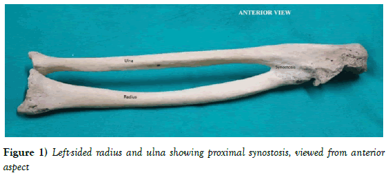
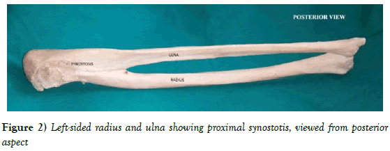
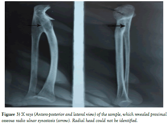
The bone specimen described in the present study involves a proximal radioulnar synostosis of the left upper limb and was obtained as an incidental finding, during routine osteology classes for undergraduate students, in the anatomy department of Murshidabad Medical College. Out of the 276 numbers of radius and ulna, only one synostotic bone was available. No gross external injury was visible. The synostotic bone was separated and considered for presentation as a case report. The measurements were taken with the help of a sliding Vernier calliper and flexible steel tape. The length of the synostosis was 7.5 cm in the left upper limb. It was a proximal synostosis. Radial head could not be identified (Figures 1-3).
Discussion
Radio-ulnar synostosis being such a rare malformation, the specimen found was significant. It is considered as an anomaly of longitudinal segmentation. Sandifort in 1973 originally described the congenital radio-ulnar synostosis, as a rare congenital anomaly. In 60% cases, it is bilateral [7,8]. However, in 9% cases it runs a familial course [9]. Again in 25% cases, there is some kind of genetic basis for the disease [10] and chromosomal malformation plays some role in this malformation. Wilkie [4] described two types of synostosis based on the radio-graphic appearance. Type 1 comprises the true radioulnar synostosis where radius and ulna are smoothly fused at their proximal ends and extends for a variable distance distally. Type 2 is characterised by congenital dislocation of the head of radius, in addition to type 1 deformity. In 1985, Clearly et al in 1985 described four separate patterns of radioulnar synostosis, radiologically [11]. Type 1 does not involve bone and is characterised by a reduced but normally appearing radial head. In Type II, bony synostosis can be seen. Type III comprises of osseous synostosis with a hypoplastic and a posteriorly dislocated radial head. In type IV, there is short osseous synostosis with an anteriorly dislocated radial head. As per Wilkie and Davenport, we had Type I radio-ulnar synostosis and according to Cleary’s classification, we had Type II synostosis. As the bone specimen under study was an incidental finding, no history of the individual was available. Surgeries should be aimed depending on the severity of the functional deficits and whether or not it is bilateral.
References
- Avadhani R, Bindhu S, Vikram, et al. Radio ulnar synostosis: A case Report. NUJHS. 2011;1.
- Dogra BB, Singh M, Malik A. Congenital proximal radioulnar synostosis. Indian J Plast Surg. 2003;36:36-8.
- Hansen OH, Anderson NO. Congenital radioulnar synostosis. Report of 37 cases. Acta Orthop Scand. 1970;41:225-30.
- Wilkie DP. Congenital radioulnar synostosis. Br J Surg. 1914;1:366-75
- Lewis WH. The development of the arm in man. Am J Anat. 1901;1:145-83
- Mital MA. Congenital radioulnar synostosis & congenital dislocation of the radial head. Orthop Clin North A. 1976;2:375-83.
- Lescau HE, Mulligan J, Williams G. Congenital radio-ulnar synostosis in an active duty soldier: case report & literature review. Mil Med. 2000;165:425-8.
- Wurapa R. Radial-ulnar synostosis. E Medicine.
- Fakoor M. Radio-ulnar synostosis in a father and his 5 year old daughter. Pak J Med Sci. 2006;22:191-3.
- Yammine K, Salon A, Pouliquen JC. Congenital radioulnar synostosis. Study of a series of 37 children & adolescents. Chir Main. 1998;17:300-8.
- Cleary JE, Omer GE. Congenital proximal radioulnar synostosis. Natural history and functional assessment. J Bone Joint Surg Am. 1985;67:539-45.
Dhur A, Mukhopadhyay B and Biswas S*
Malda Medical College, Malda, West Bengal, India.
- *Corresponding Author:
- Dr. Sharmistha Biswas
Malda Medical College, Malda, West Bengal, India
Tel: 03512 221 087
E-mail: drsharmisthabiswas@rediffmail.com
Citation: Dhur A, Mukhopadhyay B, Biswas S. Proximal radio ulnar synostosis: a case report. Int J Anat Var. 2017;10(3):47-8.
Copyright: This open-access article is distributed under the terms of the Creative Commons Attribution Non-Commercial License (CC BY-NC) (http://creativecommons.org/licenses/by-nc/4.0/), which permits reuse, distribution and reproduction of the article, provided that the original work is properly cited and the reuse is restricted to noncommercial purposes. For commercial reuse, contact reprints@pulsus.com
Abstract
Radio-ulnar synostosis constitutes one of the rare and uncommon anomalies of the upper limb. It may be congenital where there is failure of segmentation of the radius and the ulna or acquired resulting in post-traumatic synostosis. Males and females are equally affected in congenital variety and bilateral involvement is found in half of those affected. The condition may be associated with other developmental anomalies or as a part of an underlying syndrome. Clinical presentations include subtle functional limitations of the elbow flexion and extension and pain is a rare complaint. However, a careful medical history, aided by physical examination and diagnostic tool can confirm the diagnosis. Here, we are reporting a case of proximal radio-ulnar synostosis with an aim to make clinicians aware of the possible differential diagnosis and thereby successful treatment outcome.
-Keywords
Synostosis; Segmentation; Functional limitations; Radioulnar synostosis
Introduction
Synostosis is a term applied to the union of two adjacent bones of the body. Radio-ulnar synostosis is a rare anomaly resulting from the fusion of two bones of forearm, i.e., the radius and the ulna. It exists in two forms: congenital and post- traumatic; congenital one result from failure of prenatal separation of the radius and ulna, post-traumatic synostosis is also not uncommon [1]. However, the degree of union varies and the radial head may remain unaffected. Congenital radio-ulnar synostosis often occurs as part of syndromes such as Crouzon, Apert’s and Poland’s [2]. Functional limitations of the upper limb are a common presenting complaint. Although the exact etiology is not known, a genetic basis has been documented for such fusion [3]. During embryonic period the forearm lies pronated and the same position is found in almost all radio-ulnar synostosis [4]. However, the embryological basis can be explained by the fact that the humerus, radius and ulna are continuous with each other and are joined by a common perichondrium at 5 weeks of gestation [5]. By 6th week, the cartilaginous analogue of the three bones is separated by condensations of tissue. A failure of prenatal longitudinal segmentation with persistence of cartilaginous analogue between radius and ulna during 7th week of development results in persistent bridge of tissue [6]. The diagnosis of the synostosis is established radiologically. Both limbs should be simultaneously radiographed and compared. They help to detect underlying etiology such as callus formation after trauma.
Case Report
The bone specimen described in the present study involves a proximal radioulnar synostosis of the left upper limb and was obtained as an incidental finding, during routine osteology classes for undergraduate students, in the anatomy department of Murshidabad Medical College. Out of the 276 numbers of radius and ulna, only one synostotic bone was available. No gross external injury was visible. The synostotic bone was separated and considered for presentation as a case report. The measurements were taken with the help of a sliding Vernier calliper and flexible steel tape. The length of the synostosis was 7.5 cm in the left upper limb. It was a proximal synostosis. Radial head could not be identified (Figures 1-3).
Discussion
Radio-ulnar synostosis being such a rare malformation, the specimen found was significant. It is considered as an anomaly of longitudinal segmentation. Sandifort in 1973 originally described the congenital radio-ulnar synostosis, as a rare congenital anomaly. In 60% cases, it is bilateral [7,8]. However, in 9% cases it runs a familial course [9]. Again in 25% cases, there is some kind of genetic basis for the disease [10] and chromosomal malformation plays some role in this malformation. Wilkie [4] described two types of synostosis based on the radio-graphic appearance. Type 1 comprises the true radioulnar synostosis where radius and ulna are smoothly fused at their proximal ends and extends for a variable distance distally. Type 2 is characterised by congenital dislocation of the head of radius, in addition to type 1 deformity. In 1985, Clearly et al in 1985 described four separate patterns of radioulnar synostosis, radiologically [11]. Type 1 does not involve bone and is characterised by a reduced but normally appearing radial head. In Type II, bony synostosis can be seen. Type III comprises of osseous synostosis with a hypoplastic and a posteriorly dislocated radial head. In type IV, there is short osseous synostosis with an anteriorly dislocated radial head. As per Wilkie and Davenport, we had Type I radio-ulnar synostosis and according to Cleary’s classification, we had Type II synostosis. As the bone specimen under study was an incidental finding, no history of the individual was available. Surgeries should be aimed depending on the severity of the functional deficits and whether or not it is bilateral.
References
- Avadhani R, Bindhu S, Vikram, et al. Radio ulnar synostosis: A case Report. NUJHS. 2011;1.
- Dogra BB, Singh M, Malik A. Congenital proximal radioulnar synostosis. Indian J Plast Surg. 2003;36:36-8.
- Hansen OH, Anderson NO. Congenital radioulnar synostosis. Report of 37 cases. Acta Orthop Scand. 1970;41:225-30.
- Wilkie DP. Congenital radioulnar synostosis. Br J Surg. 1914;1:366-75
- Lewis WH. The development of the arm in man. Am J Anat. 1901;1:145-83
- Mital MA. Congenital radioulnar synostosis & congenital dislocation of the radial head. Orthop Clin North A. 1976;2:375-83.
- Lescau HE, Mulligan J, Williams G. Congenital radio-ulnar synostosis in an active duty soldier: case report & literature review. Mil Med. 2000;165:425-8.
- Wurapa R. Radial-ulnar synostosis. E Medicine.
- Fakoor M. Radio-ulnar synostosis in a father and his 5 year old daughter. Pak J Med Sci. 2006;22:191-3.
- Yammine K, Salon A, Pouliquen JC. Congenital radioulnar synostosis. Study of a series of 37 children & adolescents. Chir Main. 1998;17:300-8.
- Cleary JE, Omer GE. Congenital proximal radioulnar synostosis. Natural history and functional assessment. J Bone Joint Surg Am. 1985;67:539-45.


