Retrorenal appendix: An atypical position of the vermiform appendix
Received: 05-Aug-2018 Accepted Date: Aug 24, 2018; Published: 07-Sep-2018
Citation: Gandhi JA, Shinde P, Shetty T, et al. Retrorenal appendix: an atypical position of the vermifom appendix. Int J Anat Var. Sep 2018;11(3):099-100.
This open-access article is distributed under the terms of the Creative Commons Attribution Non-Commercial License (CC BY-NC) (http://creativecommons.org/licenses/by-nc/4.0/), which permits reuse, distribution and reproduction of the article, provided that the original work is properly cited and the reuse is restricted to noncommercial purposes. For commercial reuse, contact reprints@pulsus.com
Summary
Purpose: Appendicitis is one of the most common clinical conditions requiring emergency surgery. Variations in the anatomical position of vermiform appendix can result in different clinical presentations. A thorough knowledge of normal anatomy, variations in the position and corresponding radiological image of vermiform appendix is essential for radiologists and surgeons in planning clinical and surgical management for pathologies afflicting the appendix.
Methods and Results: A 55-year-old patient presented to the Emergency Department with recurrent lower abdominal pain, insidious in onset and burning in character, associated with vomiting. On physical examination, however, her abdomen was soft and non-tender. Further radiological investigations revealed appendicitis with the position of the appendix being retrorenal.
Keywords
Retrorenal appendix; Vermiform appendix
Introduction
Beginning from the first drawings of the appendix by Leonardo da Vinci in 1492 [1], the study of anatomy and embryology of the appendix has captured the attention of the surgical fraternity right up to the mid twentieth century. From the time of Sir Frederick Treves’ mention of the ‘splenic’ or post ileal appendix being the most common position of the vermiform appendix to it being challenged by Cecil Wakeley with his study claiming that this blind diverticulum was most often found in the retro-caecal location, it has been repeatedly observed that the appendix can be found in almost any position relative to the cecum [2]. There is no definite rule governing the position of the appendix. Its length varies from 2 cm to 20 cm with an average of 10 cm [3]. The base of appendix is fixed whereas the remaining part of the appendix may occupy any of the following reported positions indicated by the hour hand of the clock- i) retrocaecal (12 O’clock), ii) preileal (1 O’clock) iii) postileal (2 O’clock) iv) promonteric (3 O’clock) v) pelvic (4 O’clock) and vi) subcaecal (6 O’clock) [4-7].
Although vestigial it can be a significant source of morbidity and occasionally mortality, when inflamed. In spite of being one of the most common surgical procedures performed by the surgeon, appendicectomy is fraught with pitfalls for the unwary due to the highly variable position of the appendix.
Herein described is, to the best of our knowledge, one such never before described anatomic variation of appendix.
Case Report
A 55-year-old lady presented to the emergency department of a tertiary care centre with complaint of diffuse burning lower abdominal pain for 15 days, predominantly in the right lumbar region, which was insidious in onset, moderate in intensity, aggravated with exertion not associated with vomiting. She gave history of similar pain intermittently since the past two months relieved on medication but the present pain was not relieved by analgesics. The pain was not associated with bowel and bladder symptoms. She was a known seropositive patient (HIV 1 positive) and was on antiretroviral medications for the past 5 years. She had no other co morbidity.
On examination, general condition of the patient was fair with normal vital parameters. Per abdomen the patient was soft on palpation, no tenderness or guarding was appreciated over the abdomen. Routine investigations were within the normal range except for mildly elevated WBC count of 12,500. Chest and erect abdominal X-rays were normal. On ultrasonography of the abdomen and pelvis, the appendix could not be visualised. Considering the age of the patient, leucocytosis and her seropositive status a contrast enhanced CT scan of the abdomen and pelvis was done to rule out any finding missed on the ultrasound scan.
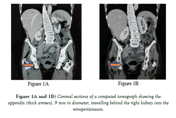
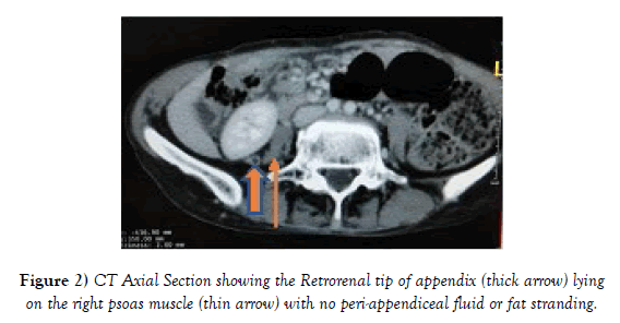
CT scan showed a blind tubular structure arising from the caecum travelling back into the retroperitoneum with its tip lying behind the right kidney. The tubular structure was confirmed as the appendix by radiologists with a diameter of 9 mm. The caecum was orthotopic in position. There was no sign of peri appendiceal fat stranding or peri appendiceal fluid. No other significant abnormality could be detected on the scan. Based on the clinical and radiological profile of the patient, a diagnosis of acute appendicitis was derived.
The notable finding on the scan was the unusual position of the appendix, a never before seen or described position of the vermiform appendix (Figure 1). As the tip of the appendix ended behind the right kidney, it has been termed as Retrorenal appendix (Figure 2). In view of a soft and non-tender abdomen, normal vital parameters and lack of signs of active inflammation of the appendix on computed tomography of the abdomen, the patient was managed conservatively with IV antibiotics and analgesics. After relief of symptoms and normalization of leucocyte count, she was discharged and planned for laparoscopic interval appendicectomy. However, the patient failed to report for follow up visits.
Discussion
Embryologically the appendix is the terminal portion of the caecal diverticulum. The base of the appendix has a constant relation with caecum, i.e. on the posteromedial aspect of the caecum 2 cm below the IC junction, but the tip can point in various directions. The position of the tip depends on many factors including peritoneal fixation, configuration of the caecum, appendiceal length, associated adhesions, and the habitus of the person but most profoundly on the changes in position and shape of the cecum during growth and development [2,8].
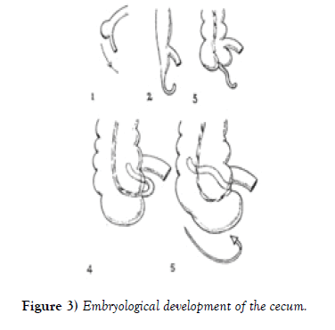
The cecum first appears as a saccular diverticulum of the midgut demarcating the large intestine from the small intestine (Figure 3). This is followed by the relative descent of the cecum and ascent of the liver with the rapid growth of the abdominal wall and ascending colon. Helicoidal torsion of the cecum, then, results in the leftward, upward and backward displacement of the appendico-caecal junction giving rise to the retrocaecal position of the appendix.
Till date majority of the described anatomical variations of the appendix have been peritoneal structures. Only 7% are retroperitoneal (or retrocolic) [9]. In this case the retrocecal appendix is seen travelling into the retroperitoneum between the paranephric fat and the Gerota’s fascia with the tip lying behind the right kidney. Hence it is termed as a retrorenal appendix.
Conclusion
In addition to expanding the literature on the anatomical variations of the vermiform appendix, this case implores surgeons to keep an open mind to the range of possible positions of the appendix when diagnosing both common and rare pathologies affecting this organ and while planning surgical interventions involving the appendix.
REFERENCES
- Pappas S. Anatomy Meets Art: Da Vinci’s Drawings. 2012.
- Wakely CPG. The position of vermiform appendix as ascertained by an analysis of 10000 cases. J Anat. 2000;67:277.
- Richmond B. The Biological Basis of Modern Surgical Practice. Elsevier, USA 2017;pp:1296-311.
- Ahmed I, Asgeirsson KS, Beckingham IJ, et al. The position of the vermiform appendix at laparoscopy. Surg radiol anat. 2007;29:165-8.
- Banerjee A, Kumar I A, Tapadur A. Morphological variations in the anatomy of caecum and appendix a cadaveric study. National j cli anat. 2005;1:30-5.
- Delic J, Savkovic A, Isakovic E. Variations in the position and point of origin of the vermiform appendix. 2005.
- Paul UK, Naushaba H, Alam MJ, et al. Length of the vermiform appendix: a post-mortem study. Bangladesh J Anat. 2000.
- Beneventano TC, Schein CJ, Jacobson HG. The Roentgen aspects of some appendiceal abnormalities. Am J Roentgenol Radium Ther Nucl Med. 1966; 96:344-60.
- Marbury WB. The retroperitoneal (retrocolic) appendix. Ann Surg. 1938;107:819-28.


