Study of formation of pelvic somatic nerves
Received: 20-Nov-2017 Accepted Date: Dec 06, 2017; Published: 11-Dec-2017
Citation: Preeti DS, Neelesh K, Vatsalaswamy P. Study of formation of pelvic somatic nerves. Int J Anat Var. 2017;10(4): 106-111.
This open-access article is distributed under the terms of the Creative Commons Attribution Non-Commercial License (CC BY-NC) (http://creativecommons.org/licenses/by-nc/4.0/), which permits reuse, distribution and reproduction of the article, provided that the original work is properly cited and the reuse is restricted to noncommercial purposes. For commercial reuse, contact reprints@pulsus.com
Abstract
Variations are very commonly found in the formation of nerves. They may be prefixed or post fixed. Variable thickness of neural roots or their absences are also commonly seen. The pelvic nerves lie along the posterior pelvic wall except obturator nerve. The obturator nerve lies along the lateral pelvic wall. Pelvic nerves are present extraperitoneally against the posterolateral walls of pelvis. The somatic nerves of the pelvis include the Lateral Femoral Cutaneous nerve, Femoral nerve, Genitofemoral nerve, Obturator nerve and the Lumbosacral trunk. Variations in the level of roots of these nerves are very commonly seen which are important for surgeons, orthopaedic surgeons and neurosurgeons during different surgeries. . Different types of variations were found in the formation as well as level of division of parietal pelvic nerves which are discussed in the study.
Keywords
Lateral femoral cutaneous nerve; Femoral nerve; Genitofemoral nerve; Obturator nerve; Lumbosacral trunk; Variations; Roots
Introduction- Nerves of the Pelvis
The nerves of pelvis are related to the posterior wall of pelvis except obturator nerve. The obturator nerve passes along the lateral wall of pelvis. These nerves lie within the pelvic cavity.
The somatic nerves of the pelvis are the Lateral Femoral Cutaneous nerve, femoral nerve, Genitofemoral nerve, Obturator nerve and the Lumbosacral trunk.
Lateral Femoral Cutaneous nerve arises from posterior divisions of L2–L3, it passes obliquely across the iliacus muscle towards anterior superior iliac spine. Then it Passes beneath the inguinal ligament and divides into an anterior and posterior branchs. The anterior branch gives cutaneous supply to the anterolateral aspect of the thigh. The posterior branch pierces the fascia lata and gives cutaneous supply to the skin of the lateral aspect of thigh from greater trochanter to middle of the thigh.
Femoral nerve is formed from the posterior divisions L2, L3, L4. It is the nerve which gives cutaneous supply to the anterior aspect of the thigh and it is one of the largest branch of the lumbar plexus. It passes through the fibres of psoas major muscle and descends between the psoas major and the iliacus then passes beneath the inguinal ligament just lateral to the femoral artery as it enters the thigh.
Inside the abdominal cavity the femoral nerve gives off muscular branches to the iliacus. It also gives two large cutaneous branches i.e. anterior cutaneous branch which gives intermediate and medial cutaneous nerves. The intermediate femoral cutaneous nerve descends along the anterior aspect of thigh to supply the skin and then contributes to the patellar plexus.
The medial femoral cutaneous nerve supplies the skin on the medial side of the thigh. The femoral nerve gives several muscular branches including the nerve to pectineus, nerve to vastus medialis, nerve to vastus lateralis, nerve to vastus intermedius, nerve to sartorius, and the large cutaneous branch i.e. saphenous nerve.
Genitofemoral nerve is formed from L1&L2 roots. It penetrates the psoas major muscle and runs inferiorly along the anterior aspect of the muscle beneath the fascia transversalis and the peritoneum, and then bifurcates into genital and femoral branches.
The genital branch reaches the spermatic cord; this nerve supplies the cremaster muscle and the skin of the scrotum and thigh. In females, it accompanies the round ligament of the uterus. The femoral branch passes beneath the inguinal ligament enters the femoral sheath, exits the sheath as well as fascia lata to supply the skin of the upper part of the anterior aspect of thigh.
Obturator nerve arises from anterior divisions of L2, L3&L4. It emerges from the medial border of the psoas major muscle it travels along the lateral wall of the lesser pelvis and then enters in to the obturator foramen. After entering in to the thigh, obturator nerve bifurcates into an anterior and posterior branch. The anterior branch passes anterior to the obturator externus, deep to the pectineus and adductor longus, and superficial to the adductor brevis. Muscular branches give supply to the adductor longus, gracilis, and adductor brevis. The posterior branch of the obturator nerve exits the anterior aspect of the obturator externus, travels beneath the adductor brevis anterior to the adductor magnus, and then it gives off muscular and articular branches. The muscular branches innervate the obturator externus, adductor magnus, and the adductor brevis [1].
Lumbosacral trunk is a thick cord which is formed from the entire ventral ramus of the L5 and the descending part of the L4. It descends obliquely over the lateral part of the sacrum into the pelvis. Then it passes above the superior gluteal vessels across the pelvic surface of the sacro-iliac joint, to join the sacral ventral rami in front of pyriformis muscle [2].
The lumbar part of lumbosacral trunk contains part of the fourth and all the fifth lumbar ventral rami of L4, L5; it appears along the medial margin of psoas major muscle, and then passes over the pelvic brim in front of the sacroiliac joint to join the first sacral ramus. The greater part of second and third sacral rami converges on the inferomedial aspect of the lumbosacral trunk in the greater sciatic foramen and they form the sciatic nerve [3].
Differences in the formation of pelvic nerves detected during spinal operations have motivated orthopaedic surgeons and anatomists to work on the variations of Lumbosacral plexus formation. Aim of this study was to find out variations in the formations of the nerves of the pelvis as well as their course, which is surgically important.
Also the purpose of this study was to describe the anatomical variations in the somatic nerves of the pelvis from their roots to the exit from the pelvic cavity.
This study was undertaken as intraoperative injury of nerves are common risks repeatedly described in orthopaedic and gynaecological surgeries and hence one should be aware of the variations of pelvic nerves.
Materials and Methods
40 pelvises were dissected from the department of Anatomy of Dr. D. Y. Patil Medical College, Pimpri, Pune. These cadavers were embalmed with 10 per cent formalin and fixed. They was labeled from 1-40 out of 40 cadavers 35 were male cadavers and 5 were female cadavers. Left and right side were also labeled. Pelvis was separated from the cadaver and sagittal section of the pelvis was taken, by cutting the sacrum in the midline. Dissection was carried out following the procedure as per Cunningham’s manual [4].
The steps of dissection were as follows:
• Dissected cadavers were cut at the level of twelfth thoracic vertebra.
• Sagital section of this cut pelvis was taken in the midline.
• The specimen was labelled with number & side.
• Psoas major muscle was removed in piecemeal to expose the branches of lumbar plexus which are confined to the pelvis i.e. Lateral Femoral Cutaneous nerve, Genitofemoral nerve, femoral nerve, obturator nerve and lumbosacral trunk.
• All the nerves and their variations were noted with the side.
• Photographs of variations were taken.
Observations
Variations of the following nerves of the pelvis were recorded according to right and left sides (Tables 1-5).
| Name | Variations if any | Sp. No. | Side | Sex | Percentage | |
|---|---|---|---|---|---|---|
| Rt | Lt | |||||
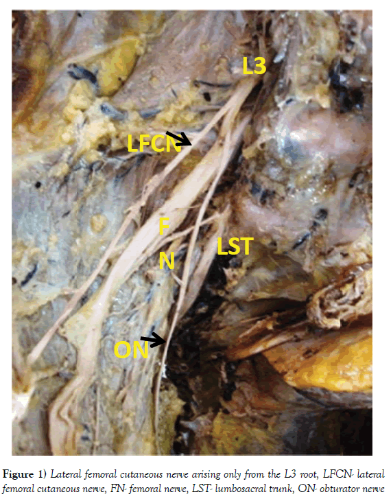
| Lateral femoral cutaneus nerve | Only from the L 3 root (Figure 1) | 5 15 20 26 31 |
√ - - √ √ |
√ √ √ - - |
M F M M M |
12.5% |
| Prefixed from the L1 and L 2 | 7 9 10 16 |
- - √ √ |
√ √ - √ |
M M M M |
10% | |
| Accessory LFCN | 25 30 |
√ - |
- √ |
M M |
5% | |
Table 1: Lateral femoral cutaneous nerve
| Name | Variations if any | Sp. No. | Side | Sex | Percentage | |
|---|---|---|---|---|---|---|
| Rt | Lt | |||||
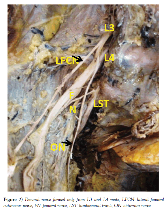
| Femoral nerve | Contribution only from L 3 and L 4 (Figure 2) | 5 | √ | √ | M | 2.5% |
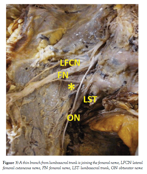
| A thin branch from the lumbosacral trunk joining the femoral nerve (Figure 3) | 25 30 |
√ - |
- √ |
M M |
5% | |
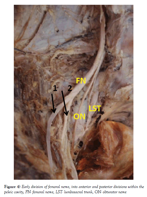
| Early division within the pelvis in to two branches (Figure 4) | 6 13 27 |
√ - - |
√ √ |
F M M |
7.5% | |
Table 2: Femoral nerve
| Name | Variations if any | Sp. No. | Side | Sex | Percentage | |
|---|---|---|---|---|---|---|
| Rt | Lt | |||||
| Genito femoral nerve | Premature bifurcation into genital and femoral | 4 17 |
√ √ |
- - |
M M |
5% |
| Genital and femoral arising separately from the lumbar plexus | 3 18 28 |
√ √ √ |
- - - |
M M M |
7.5% | |
Table 3: Genito femoral nerve
| Name | Variations if any | Sp. No. | Side | Sex | Percentage | |||
|---|---|---|---|---|---|---|---|---|
| Rt | Lt | |||||||
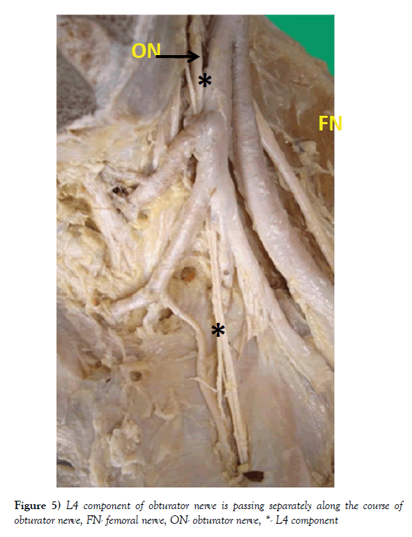
| Obturator nerve | Extra branch from the L 4 accompanying the obturator nerve (Figure 5) | 11 12 35 |
√ √ √ |
- √ - |
M F F |
7.5% | ||
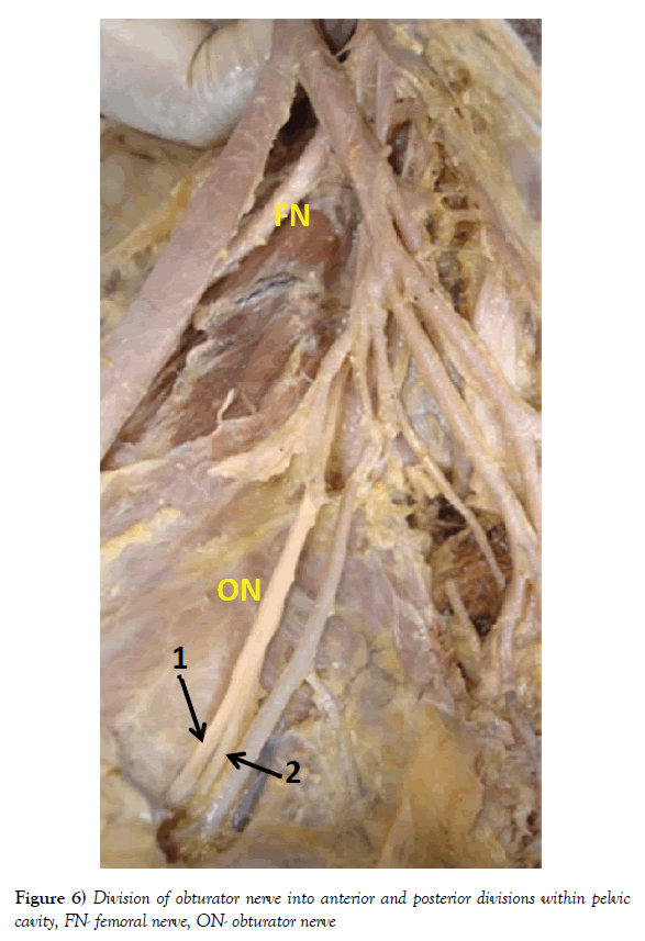
| Division of obturator nerve in to anterior and posterior divisions within pelvic cavity (Figure 6) |
4 | √ | - | M | 2.5% | |||
| Accessory obturator nerve | 6 | √ | √ | F | 2.5% | |||
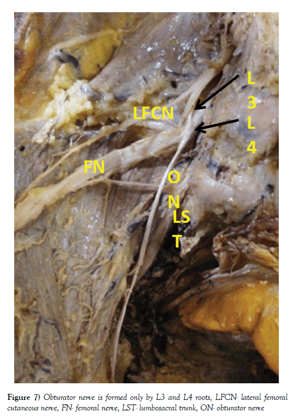
| Contribution only from L3 , L4 (Figure 7) |
5 | √ | √ | M | 2.5% | |||
Table 4: Obturator nerve
| Name | Variations if any | Sp. No. | Side | Sex | Percentage | |
|---|---|---|---|---|---|---|
| Rt | Lt | |||||
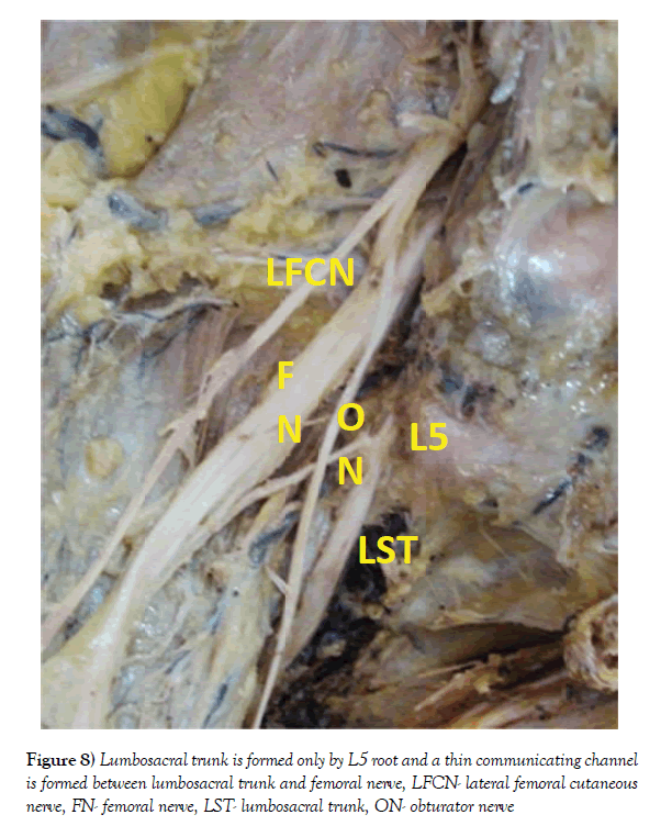
| Lumbosacral trunk | No contribution from L 4 (Figure 8) | 5 | √ | √ | M | 2.5% |
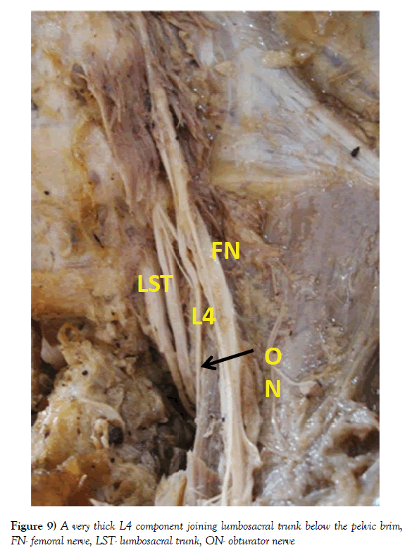
| Very thick L 4 component (Figure 9) |
2 22 24 29 34 |
√ √ √ √ √ |
√ √ √ √ √ |
M F M M M |
12.5% | |
| Very thin L 4 component | 1 8 19 21 23 |
√ √ √ √ √ |
√ √ √ √ √ |
M M M M M |
12.5% | |
| L 4 joins above pelvic brim | 22 29 34 |
√ √ √ |
√ √ √ |
M F M |
7.5% | |
| L 4 component joins below pelvic brim | 21 23 |
√ √ |
√ √ |
M M |
5% | |
Table 5: Lumbosacral trunk
Discussion
Pelvic (somatic) nerves i.e. the lateral femoral cutaneous nerve, femoral nerve, genitofemoral nerve, obturator nerve and lumbosacral trunk were studied for their formation and variations in their formation and the findings of the present study were compared with the previous studies also (Figures 1-9).
Lateral Femoral Cutaneous nerve
Anloague found variation in the formation of the lateral femoral cutaneous nerve in about 17.6% of the cases. The lateral femoral cutaneous nerve normally arose from the posterior divisions of the L2 and L3 roots, in 4 cases, it was arising from the L1 and L2 nerve roots and in one case it was having its origin only from the L2 root. Another variation included a bifurcation of the lateral femoral nerve within the pelvic cavity before its exit near the anterior superior iliac spine; bifurcations normally occur after the nerve exits the pelvic cavity [5].
Bergman found that sometimes Lateral Femoral cutaneous nerve was arising from the femoral nerve, also sometimes it was absent, and it was also found arising from the L1 and L2 or from L3 and L4 roots [6].
Present study showed the formation of Lateral Femoral Cutaneous nerve only from the L3 root in 8.6% cases. The Lateral Femoral Cutaneous nerve was also arising from the L1 and L2 roots which were similar to the finding of Erbil [7]. Also in 3.8% cases the accessory Lateral Femoral Cutaneous nerve was found.
Femoral nerve
Anloague et al. observed the formation of two divisions of femoral nerve; in most cadavers, this occurred within the substance of the psoas major muscle. One case showed medial and lateral bifurcation of the nerve within psoas major musculature and an intermediate connection between the two slips of the nerves [5]. Uzmansel et al. observed the femoral nerve receiving a slender branch from the Lateral Femoral Cutaneous nerve [8]. Erbil observed the formation of femoral nerve taking place from the anterior rami of L2, L3, L4, and L5 [7]. Jamieson found the accessory femoral nerve which he called as the accessory anterior crural nerve [9].
Bergman found numerous variations in the formation of the femoral nerve, sometimes it was formed by L1, L2, L3, L4 sometimes it had contribution from the T12 root also. In few cases the femoral nerve had the root value as L1, L2, L3, L4, L5 [6]. The present study showed formation of the femoral nerve only by the L3 and L4 roots in 2.9% cases. The present study also showed a small branch from the Lumbosacral Trunk joining the femoral nerve in 3.1% cases. The femoral nerve was dividing in to two branches, 3.2 cm above the midpoint of inguinal ligament which was similar to the finding of Das [10].
Genitofemoral nerve
Anloague found variations in the formation of genitofemoral nerve in 47.1% of cases. Normally, it bifurcates into its terminal, genital and femoral branches midway along the anterior surface of the psoas major muscle. The most common variation was seen in 26.5% cases and included a split of the genitofemoral nerve into the genital and femoral branches within the substance of the psoas major muscle. Bergman found bifurcation of genitofemoral nerve at the upper part of the anterior surface of the psoas major muscle in 20.6% cases [5]. Uzmansel et al. found the formation of Genitofemoral nerve from the ventral root of the L2 only. Sometimes it was formed from only the L3 component [8].
Bergman observed the absence of genital or femoral branches of the genitofemoral nerve. Also in 80% of cases the genitofemoral nerve was not dividing in to its two branches i.e. genital and femoral nerves. The variations in the formation of the genitofemoral nerve were found like formation from the L1, L2 or L2, L3 or from the L1, L2, and L3 [6]. The present study showed the early bifurcation of the genitofemoral nerve in to its two branches in 2.9% of cases. And the two branches, genital and femoral of the genitofemoral were arising separately from the lumbar plexus in 4.5% of cases.
Obturator nerve
Erbil observed variable formation of the obturator nerve, it was formed from the L1, L2, L3 roots or sometimes it was having additional root from the L1 or from L5 [7].
Bergman found its formation from L1, L3, L4, L5 and the contribution from the L2 was missing [6].
Akkaya found the accessory obturator nerve in 12.5% cases. It’s formation was from the L2, L3, and L4 or sometimes from the L2 and L3 [11]. In present study the obturator nerve was formed from L4 and L5 in 3% cases. A small branch from the L4 was accompanying the obturator nerve in 4.3% cases. The present study also showed the presence of accessory obturator nerve in 3.1% cases. It was bilateral. In 4.3% of cases it was formed only from L3 and L4 roots.
Obturator nerve lie along the pelvic wall hence it is subject to possible damage when it is blocked for partial denervation of adductor muscles or partial denervation of hip joint. It also may get damaged in extensive removal of lymph nodes of the pelvis, as in the Wertheim’s operation [3].
Lumbosacral trunk
Bergman studied variations in the formation of lumbosacral trunk , like contribution from the L3 or L4. Sometimes the contribution from the L4 was absent, also the L4 component called as nervi furcalis , was very thin or sometimes it was very thick [6].
In the present study there was no contribution from L4 in the formation of the lumbosacral trunk in 2.9% cases.
In the present study Nervi furcalis was thin in 17.3% cases, while it was very thick which were similar to the finding of Bergman.
Also in the present study the L4 component was joining the lumbosacral trunk above the pelvic brim in 11.5% cases while the L4 component was joining it below the pelvic brim in 8.7% cases.
Meralgia paresthetica is a condition which includes numbness, paresthesia and pain in the anterolateral aspect of thigh, which may result from an entrapment neuropathy or a neuroma of the lateral femoral cutaneous nerve [12]. Hence lateral femoral cutaneous nerve was studied. The congenital anomalies of the variations of lumbosacral nerves have frequently observed in the past as operative finding during surgery of vertebral column and now a day’s diagnosed preoperatively with more frequency. All these different types of formations of pelvic nerves are very important for surgeons and anaesthetists.
Embryological explanation –In case of the nerves supplying the limb buds, to form the cervico-brachial and lumbosacral plexuses the anterior primary rami are joined by connecting loops. Posterior divisions of the trunks of these plexuses supply the extensor muscles and anterior divisions supply the flexor muscles. So when these primary rami join to form the loops they may fuse to the previous roots to form prefixed plexuses or they may fuse with the next roots to form post fixed plexuses and it leads into variations in the formation of nerves [13].
REFERENCES
- Henry Gray. The Anatomical Basis of Clinical Practice. Elsevier Churchill Livingstone. 2005;1360-5.
- Last’s Anatomy: Regional and Applied. RMH Mcminn, Churchill Livingstone. 1994;394-8.
- Henry Hollinshead W. Anatomy for surgeons. Harper & row publishers. PP: 665-70.
- Romanes GJ. Cunningham’s manual of practical anatomy. Oxford medical publications. 1986;2:232-8.
- Philip A Anloague. Anatomical Variations of the Lumbar Plexus: A Descriptive Anatomy Study with Proposed Clinical Implications. J Manual Manipulative Ther. 1998; 17:4.
- Ronald A Bergman. Variations of Connections between Lumbar and Sacral Plexuses. Folis Morphol. 1981;40:271-9.
- Erbil KM. Examination of the variations of the lateral femoral cutaneous nerves: Report of two cases. Anat Sci Inc. 2002;77:247-9.
- Deniz Uzmansel. Multiple variations of the nerves arising from the lumbar plexus. Neuroanatomy. 2006;5:37-39.
- Jamieson EB. The origin and localization of the accessory obturator nerve. Nauchni Tr Vissh Med Inst Sofiia. 1903; 39:1-12.
- Srijit Das. Anomalous branching pattern of the femoral nerve. Acta Medica. 2007;50:245-6.
- Akkaya T. Detailed anatomy of accessory obturator nerve blockade. Minerva Anestesiol. 2008;74:119-22.
- Matejcik V. Anatomical variations of lumbosacral plexus. Surg Radiol Anat.
- WJ Hamilton, HW Mossman. Hamilton, Boyd and Mossman’s Human Embryology: prenatal development of form and function. Williams and Wilkins. 1972; PP: 491.


