Sural nerve with no contribution from tibial nerve – a rare case
Karabi BARAL, Sharmistha Biswas*, Deepraj Mitra and Soma Saha
Department of Anatomy, N.R.S. Medical College, Kolkata, India
- *Corresponding Author:
- Dr. Sharmistha Biswas
Associate Professor, Department of Anatomy, N.R.S. Medical College, Kolkata 14, 700091, India
Tel: +91 (990) 3408977
E-mail: drsharmisthabiswas@rediffmail.com
Date of Received: August 6th, 2011
Date of Accepted: August 2nd, 2012
Published Online: December 25th, 2012
© Int J Anat Var (IJAV). 2012; 5: 130–131.
[ft_below_content] =>Keywords
sural nerve, peripheral nerve transplantation, cutaneous nerve variation in leg
Introduction
Sural nerve (SN) is a sensory nerve which supplies the skin of the posterolateral aspect of the distal one third of the leg, lateral malleolus, along the lateral side of the foot and little toe. The lateral sural cutaneous nerve (LSCN) arises from the common peroneal nerve (also called common fibular nerve or CFN), supplies the skin over the upper part of the lateral and posterolateral aspect of the leg. It often gives a communicating branch called peroneal communicating branch to the medial sural cutaneous nerve (MSCN) from tibial nerve (TN) and thus form sural nerve (SN) [1]. According to standard textbooks, CFN gives rise to two cutaneous branches, often from a common trunk –they are lateral sural nerve and sural communicating nerve (SCN). SCN arises near the head of fibula and crosses the lateral head of gastrocnemius muscle to join sural nerve. SCN may descend separately as far as the heel. According to some authors, SCN is also called LSCN [2,3].
Clinically, SN is widely used for both diagnostic purpose (biopsy and nerve conduction velocity study) and therapeutic purpose (nerve grafting and surgical reconstruction) [4].
Case Report
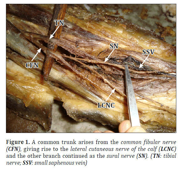
During routine cadaveric dissection for teaching purpose at the Department of Anatomy, NRS Medical College, Kolkata, a rare variation was detected in formation of SN in the left lower limb of a 67-year-old male cadaver. In this case, the SN was continuation of sural communicating branch of CFN. There was no contribution of MSCN branch of TN in the popliteal fossa or below. From the CFN, a common trunk arose, giving rise to the lateral cutaneous nerve of the calf and the other branch continued as the SN (Figure 1).
The sural nerve coursed downwards and medially to lie on the lateral side of small saphenous vein (SSV) (Figure 1). At the distal one third of leg, it crossed the SSV superficially, descended just medial to the SSV and then passed posterior to the lateral malleolus. It terminated by supplying the skin of lateral border of the foot and little toe.
No variation was found in the branching pattern of tibial nerve of the left lower limb except the absence of MSCN. The branches and courses of the nerves of right side were as usual.
Discussion
In an anatomical study of formation of SN, it was observed that 67.1% SN were formed by union of MSCN and LSCN, in 32.2% of cases SN was direct continuation of MSCN, in 0.7% of cases SN was formed by the union of MSCN and a different branch of CFN [5]. Pyun et al., observed that SN was formed by union of MSCN and LSCN in 76.9% of cases, SN was direct continuation of MSCN in 15.4% cases, and there was no communication between MSCN and LSCN in 7.7% cases [6]. Another study revealed that in 80% of cases SN was formed by union of MSCN and LSCN, and it was a continuation of MSCN in 20% of cases [7].
SN was found to be predominantly formed by MSCN and LSCN (in 87.5% of cases); and in 100% of cases, SN was found to pass along the lateral side of SSV [8]. Bilateral symmetrical origin of SN was noted to be 60.8% by Shankar et al. [9].
Ours is a rare case where SN was formed with no contribution from MSCN, it was a unilateral variation, found only on the left side. After extensive review of literature, we found only one such case report where CFN gave rise to a common trunk which, in turn divided into LSCN and SN. The MSCN, which arises from TN, did not involve in the formation of SN [10]. They found that the SN was lying lateral to the SSV throughout its course, but in our case the SN crossed the SSV superficially to lie just medial to it in the lower third of leg.
Clinically, SN is used in sensory nerve grafting for therapeutic purposes because of its long course; it is also used in nerve conduction velocity studies for diagnostic purpose [4]. Therefore, knowledge about variation of its pattern of formation and course is important for the above surgical and diagnostic procedures. Also, possibility of anatomical variation should be considered in explaining different clinical findings, like sural nerve entrapment, disproportionate and low–amplitude sural sensory nerve action potential observed during neurological evaluation [6].
Conclusion
We report a rare case of left sided variation in the formation of SN, which arose as a branch from CFN and did not receive any contribution from MSCN, a branch of TN. It initially coursed lateral to the SSV, but in distal third of leg, it passed superficial to the vein to lie on its medial side. This finding is of great clinical significance. Since SN is widely used in biopsy and as autograft in peripheral nerve transplantation, as well as in other procedures, awareness about variation in the formation and course of the nerve has immense value to the clinicians.
References
- Hollinshead WH. Anatomy for Surgeons. Vol 3. New York, Harper & Row. 1966; 768.
- Standring S, ed. Gray’s Anatomy. The Anatomical Basis of Clinical Practice. 40th Ed., Edinburg, Churchil- Livingstone. 2008; 1427.
- Moore KL, Dally A. Clinically Oriented Anatomy. 5th Ed., Philadelphia, Lippincott, Williams & Wilkins. 635.
- Kim CH, Jung H Y, Kim M O, Lee C J. The relative contributions of medial sural and peroneal communicating nerves to the sural nerve. Yonsei Med J. 2008; 47: 415–422.
- Mahakkanukrauh P, Chomsung R. Anatomical variations of the sural nerve. Clin Anat. 2002; 15: 263–266.
- Pyun SB, Kwon HK. The effect of anatomical variation of sural nerve on nerve conduction studies. Am J Phys Med Rehabil. 2008; 87: 438–442.
- Ortiguela ME, Wood MB, Cahill DR. Anatomy of the sural nerve complex. J Hand Surg Am. 1987; 12: 1119–1123.
- Eid EM, Hegazy AM. Anatomical variations of the human sural nerve and its role in clinical and surgical procedures. Clin Anat. 2011; 24: 237–245.
- Shankar N, Selvam R P, Dhanpal N, Reddy R, Alapati A. Anatomical variations of sural nerve in the leg: a fetal study. Neurol India. 2010; 58: 24–28.
- Sankar DK, Bhanu SP, Susan PJ, Gajendra K. Variant formation of sural nerve and its distribution at the dorsum of the foot. Int J Anat Var (IJAV). 2009; 2: 33–34.
Karabi BARAL, Sharmistha Biswas*, Deepraj Mitra and Soma Saha
Department of Anatomy, N.R.S. Medical College, Kolkata, India
- *Corresponding Author:
- Dr. Sharmistha Biswas
Associate Professor, Department of Anatomy, N.R.S. Medical College, Kolkata 14, 700091, India
Tel: +91 (990) 3408977
E-mail: drsharmisthabiswas@rediffmail.com
Date of Received: August 6th, 2011
Date of Accepted: August 2nd, 2012
Published Online: December 25th, 2012
© Int J Anat Var (IJAV). 2012; 5: 130–131.
Abstract
A rare case of left sided variation in the formation of sural nerve is reported. Here, it arose as a branch from common peroneal nerve and did not receive any contribution from medial sural cutaneous nerve, a branch of tibial nerve. It initially coursed lateral to the small saphenous vein, but in distal third of leg, it passed superficial to the vein to lie on its medial side. Since sural nerve is widely used in biopsy and as autograft in peripheral nerve transplantation, as well as in other procedures, awareness about variation in the formation and course of the nerve has immense value to the clinicians.
-Keywords
sural nerve, peripheral nerve transplantation, cutaneous nerve variation in leg
Introduction
Sural nerve (SN) is a sensory nerve which supplies the skin of the posterolateral aspect of the distal one third of the leg, lateral malleolus, along the lateral side of the foot and little toe. The lateral sural cutaneous nerve (LSCN) arises from the common peroneal nerve (also called common fibular nerve or CFN), supplies the skin over the upper part of the lateral and posterolateral aspect of the leg. It often gives a communicating branch called peroneal communicating branch to the medial sural cutaneous nerve (MSCN) from tibial nerve (TN) and thus form sural nerve (SN) [1]. According to standard textbooks, CFN gives rise to two cutaneous branches, often from a common trunk –they are lateral sural nerve and sural communicating nerve (SCN). SCN arises near the head of fibula and crosses the lateral head of gastrocnemius muscle to join sural nerve. SCN may descend separately as far as the heel. According to some authors, SCN is also called LSCN [2,3].
Clinically, SN is widely used for both diagnostic purpose (biopsy and nerve conduction velocity study) and therapeutic purpose (nerve grafting and surgical reconstruction) [4].
Case Report
During routine cadaveric dissection for teaching purpose at the Department of Anatomy, NRS Medical College, Kolkata, a rare variation was detected in formation of SN in the left lower limb of a 67-year-old male cadaver. In this case, the SN was continuation of sural communicating branch of CFN. There was no contribution of MSCN branch of TN in the popliteal fossa or below. From the CFN, a common trunk arose, giving rise to the lateral cutaneous nerve of the calf and the other branch continued as the SN (Figure 1).
The sural nerve coursed downwards and medially to lie on the lateral side of small saphenous vein (SSV) (Figure 1). At the distal one third of leg, it crossed the SSV superficially, descended just medial to the SSV and then passed posterior to the lateral malleolus. It terminated by supplying the skin of lateral border of the foot and little toe.
No variation was found in the branching pattern of tibial nerve of the left lower limb except the absence of MSCN. The branches and courses of the nerves of right side were as usual.
Discussion
In an anatomical study of formation of SN, it was observed that 67.1% SN were formed by union of MSCN and LSCN, in 32.2% of cases SN was direct continuation of MSCN, in 0.7% of cases SN was formed by the union of MSCN and a different branch of CFN [5]. Pyun et al., observed that SN was formed by union of MSCN and LSCN in 76.9% of cases, SN was direct continuation of MSCN in 15.4% cases, and there was no communication between MSCN and LSCN in 7.7% cases [6]. Another study revealed that in 80% of cases SN was formed by union of MSCN and LSCN, and it was a continuation of MSCN in 20% of cases [7].
SN was found to be predominantly formed by MSCN and LSCN (in 87.5% of cases); and in 100% of cases, SN was found to pass along the lateral side of SSV [8]. Bilateral symmetrical origin of SN was noted to be 60.8% by Shankar et al. [9].
Ours is a rare case where SN was formed with no contribution from MSCN, it was a unilateral variation, found only on the left side. After extensive review of literature, we found only one such case report where CFN gave rise to a common trunk which, in turn divided into LSCN and SN. The MSCN, which arises from TN, did not involve in the formation of SN [10]. They found that the SN was lying lateral to the SSV throughout its course, but in our case the SN crossed the SSV superficially to lie just medial to it in the lower third of leg.
Clinically, SN is used in sensory nerve grafting for therapeutic purposes because of its long course; it is also used in nerve conduction velocity studies for diagnostic purpose [4]. Therefore, knowledge about variation of its pattern of formation and course is important for the above surgical and diagnostic procedures. Also, possibility of anatomical variation should be considered in explaining different clinical findings, like sural nerve entrapment, disproportionate and low–amplitude sural sensory nerve action potential observed during neurological evaluation [6].
Conclusion
We report a rare case of left sided variation in the formation of SN, which arose as a branch from CFN and did not receive any contribution from MSCN, a branch of TN. It initially coursed lateral to the SSV, but in distal third of leg, it passed superficial to the vein to lie on its medial side. This finding is of great clinical significance. Since SN is widely used in biopsy and as autograft in peripheral nerve transplantation, as well as in other procedures, awareness about variation in the formation and course of the nerve has immense value to the clinicians.
References
- Hollinshead WH. Anatomy for Surgeons. Vol 3. New York, Harper & Row. 1966; 768.
- Standring S, ed. Gray’s Anatomy. The Anatomical Basis of Clinical Practice. 40th Ed., Edinburg, Churchil- Livingstone. 2008; 1427.
- Moore KL, Dally A. Clinically Oriented Anatomy. 5th Ed., Philadelphia, Lippincott, Williams & Wilkins. 635.
- Kim CH, Jung H Y, Kim M O, Lee C J. The relative contributions of medial sural and peroneal communicating nerves to the sural nerve. Yonsei Med J. 2008; 47: 415–422.
- Mahakkanukrauh P, Chomsung R. Anatomical variations of the sural nerve. Clin Anat. 2002; 15: 263–266.
- Pyun SB, Kwon HK. The effect of anatomical variation of sural nerve on nerve conduction studies. Am J Phys Med Rehabil. 2008; 87: 438–442.
- Ortiguela ME, Wood MB, Cahill DR. Anatomy of the sural nerve complex. J Hand Surg Am. 1987; 12: 1119–1123.
- Eid EM, Hegazy AM. Anatomical variations of the human sural nerve and its role in clinical and surgical procedures. Clin Anat. 2011; 24: 237–245.
- Shankar N, Selvam R P, Dhanpal N, Reddy R, Alapati A. Anatomical variations of sural nerve in the leg: a fetal study. Neurol India. 2010; 58: 24–28.
- Sankar DK, Bhanu SP, Susan PJ, Gajendra K. Variant formation of sural nerve and its distribution at the dorsum of the foot. Int J Anat Var (IJAV). 2009; 2: 33–34.


