Tendon variations of extensor digitorum and abductor pollicis longus muscles
Necdet Kocabiyik1*, Ilkan Tatar2, Bulent Yalcin1, Fatih Yazar1 and Hasan Ozan1
1Gulhane Military Medical Academy, Department of Anatomy, Ankara, Turkey
2Hacettepe University School of Medicine, Department of Anatomy, Ankara, Turkey
- *Corresponding Author:
- Necdet Kocabiyik, MD
Associate Professor of Anatomy, Gulhane Military Medical Academy (GATA), Department of Anatomy, Etlik, 06018, Ankara, Turkey
Tel: +90 312 3043508
Fax: +90 312 3042150
E-mail: nkocabiyik@gata.edu.tr
Date of Received: January 6th, 2009
Date of Accepted: May 24th, 2009
Published Online: May 26th, 2009
© IJAV. 2009; 2: 54–56.
[ft_below_content] =>Keywords
variation, extensor tendon, extensor digitorum muscle, abductor pollicis longus muscle
Introduction
Muscle and tendon variations in hand and wrist are not rare; thus their type must be well known in practicing hand surgery. Abductor pollicis longus (APL) and extensor digitorum (ED) are known to exhibit different variations with respect to their attachments. Various studies have reported splitting of the APL and ED [1-8].
The ED has one origin and a common muscle belly that splits into three or four sections that continue as tendons to the extensor hood on the dorsum of the fingers. The anatomic variations, arrangements and prevalence of these tendons have been documented in clinical and anatomic studies [9,10].
The APL originates from the radius, interosseous membrane and ulna on the dorsum of the forearm. Its tendon travels with the extensor pollicis brevis in the first dorsal compartment. Its primary insertion is into the radial side of the base of the first metacarpal bone. There are multiple additional slips of insertion including the trapezium, dorsal aponeurosis, thenar musculature (particularly abductor pollicis brevis) and flexor retinaculum. It abducts not only the thumb, but also the wrist, and is innervated by the posterior interosseous nerve [10].
The hand, with respect to its function is one of the special organs of human body, is the most frequently injured part of the body, and complete knowledge is extremely important in hand examination, treatment and reconstruction, including orthopedic procedures as tendon transfers. Anatomical variations of the extrinsic extensor tendons are frequent and knowledge is important when assessing the traumatized or diseased hand.
Case Report
Tendon variations of ED and APL muscles were observed in a 65-year-old male formalin-fixed cadaver at the Gulhane Military Medical Academy Anatomy Dissection Laboratory. Right and left forearms were dissected in a standard manner.
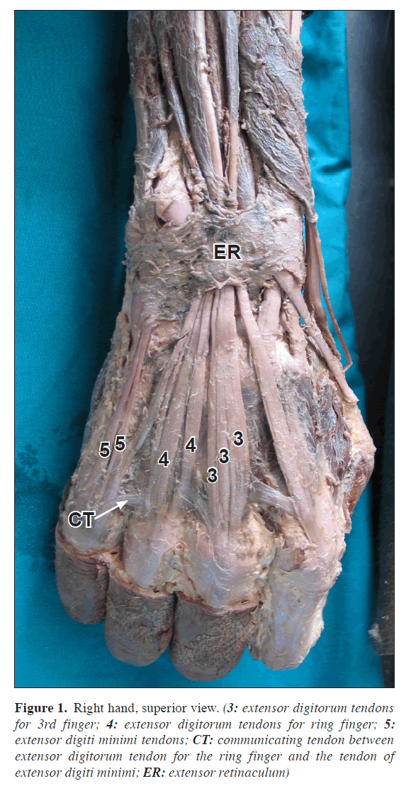
In the right forearm, the ED muscle had a tripled tendon for the 3rd finger and a doubled tendon for the ring finger. The extensor digiti minimi (EDM) also had a doubled tendon. The additional tendons began under the external retinaculum and ended at the dorsal aponeurosis; the length of these tendons were 18 cm. The intertendinous connections between the tendons of ED were in normal locations (Figure 1).
Figure 1. Right hand, superior view. (3: extensor digitorum tendons for 3rd finger; 4: extensor digitorum tendons for ring finger; 5: extensor digiti minimi tendons; CT: communicating tendon between extensor digitorum tendon for the ring finger and the tendon of extensor digiti minimi; ER: extensor retinaculum)
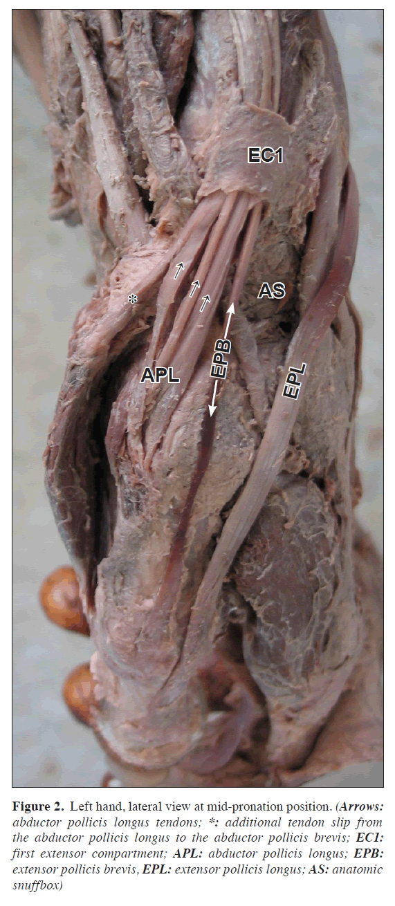
In the left forearm, the APL muscle had a tripled tendon. A thin additional tendon slip from the abductor pollicis longus was inserted to the abductor pollicis brevis. The tendons originated 5 cm proximal to the extensor retinaculum and traversed a course deep to the extensor retinaculum within the first compartment of this retinaculum. The length of these tendons was 13 cm. All tendons inserted seperatelly at the base of the first metacarpal bone. The anatomical features of extensor pollicis longus and brevis muscles were normal (Figure 2).
Figure 2. Left hand, lateral view at mid-pronation position. (Arrows: abductor pollicis longus tendons; *: additional tendon slip from the abductor pollicis longus to the abductor pollicis brevis; EC1: first extensor compartment; APL: abductor pollicis longus; EPB: extensor pollicis brevis, EPL: extensor pollicis longus; AS: anatomic snuffbox)
Discussion
The anatomic variations, arrangements and prevalence of the ED tendons have been documented in clinical and anatomic studies [2,9]. In addition to these studies there are several different studies about these extensor tendons [2,3,5,7]. Of these studies, el-Badawi et al. studied a large number of cases. In their studies, one hundred eighty-one dissected hands were examined for the pattern of extensor tendons on the dorsum of the hand [3]. ED often had multiple tendons for the middle and ring fingers. Its contribution to the little finger was usually by a bifurcating tendon common with that of the ring finger. The index finger always received a single tendon. Intertendinous connections between the various tendons of the ED were variable but were most frequent between ring and middle fingers. Extensor digiti minimi had two tendons in most cases. It was always linked to ED either by receiving one or part of its tendon or by an intertendinous connection.
Similar to above-mentioned study, our case has also multipl extensor tendons related to the middle finger; also EDM had two tendons in the same case. In another study, Seradge et al. found an anatomic variation of the EDM. The EDM had three tendon slips; two slips to the little finger and one to the ring finger’s metacarpophalangeal joint [5]. However, in our study two separate tendons were inserted to the little finger.
Fabrizio and Clemente reported a variation in the organization of the APL, bilaterally in 15 of the 50 specimens. This variation in the arrangement also brought about a retinacular-like tunnel that encased the tendons of the extensor carpi radialis longus and the extensor carpi radialis brevis muscles [4]. Sarikcioglu et al. reported an APL consisting of seven tendon slips in one case. The medial two inserted into the abductor pollicis brevis, the other five inserted into the base of the first metacarpal bone. In the right side of the same case, the abductor pollicis longus consisted of three bellies [6].
Martinez and Omer reported that bilateral subluxation of the trapeziometacarpal joint was related to an abnormal insertion of the APL tendon. They found that the APL tendon had four slips, all inserted into the fascia of the abductor pollicis brevis muscle, distal and palmar to the trapeziometacarpal joint [1].
In our study, the abductor pollicis longus muscle had a tripled tendon. The anatomical characteristics of extensor pollicis longus and brevis muscles were normal in left forearm.
The complexity and intricacy of hand function can only be revealed by understanding its anatomy. Extensor muscles have a relatively consistent architecture; however, they may have notable anatomic variations in their tendons, particularly on the ulnar side of the hand. Variations in the anatomy of the first extensor compartment have been associated with the development of de Quervain’s disease [11,12]. This disease is caused by stenosing tenosynovitis of the first dorsal compartment of the wrist, which includes the tendons of the APL and the extensor pollicis brevis (EPB). Patients usually complain of pain at the dorsolateral aspect of the wrist, reflecting towards the thumb or lateral forearm, or both [13]. Melling et al., and Ippolito et al. suggested that the number, thickness and length of the accessory tendons of APL and EPB might have an important function in the development of de Quervain’s disease [14,15]. Successful tenosynovectomy in the treatment of de Quervain disease requires paying special attention to accessory tendons of the extensor pollicis brevis and abductor pollicis longus, branching of the tendons, and the presence of an atypical septum in the first compartment.
As mentioned in the above studies, accessory tendons as in our case may be of importance in many clinical and surgical procedures.
References
- Martinez R, Omer GE Jr. Bilateral subluxation of the base of the thumb secondary to an unusual abductor pollicis longus insertion: a case report. J Hand Surg [Am]. 1985; 10: 396–399.
- von Schroeder HP, Botte MJ. Anatomy of the extensor tendons of the fingers: variations and multiplicity. J Hand Surg [Am]. 1995; 20: 27–34.
- el-Badawi MG, Butt MM, al-Zuhair AG, Fadel RA. Extensor tendons of the fingers: arrangement and variations-II. Clin Anat. 1995; 8: 391–398.
- Fabrizio PA, Clemente FR. A variation in the organization of abductor pollicis longus. Clin Anat. 1996; 9: 371–375.
- Seradge H, Tian W, Baer C. Anatomic variation of the tendons to the ring and little fingers: a cadaver dissection study. Am J Orthop. 1999; 28: 399–401.
- Sarikcioglu L, Yildirim FB. Bilateral abductor pollicis longus muscle variation. Case report and review of the literature. Morphologie. 2004; 88: 160–163.
- Zilber S, Oberlin C. Anatomical variations of the extensor tendons to the fingers over the dorsum of the hand: a study of 50 hands and a review of the literature. Plast Reconstr Surg. 2004; 113: 214–221.
- Paul S, Das S. Variant abductor pollicis longus muscle: a case report. Acta Medica (Hradec Kralove). 2007; 50: 213–215.
- Mestdagh H, Bailleul JP, Vilette B, Bocquet F, Depreux R. Organization of the extensor complex of the digits. Anat Clin. 1985; 7: 49–53.
- von Schroeder HP, Botte MJ. Anatomy and functional significance of the long extensors to the fingers and thumb. Clin Orthop Relat Res. 2001; 383: 74–83.
- Gonzalez MH, Sohlberg R, Brown A, Weinzweig N. The first dorsal extensor compartment: an anatomic study. J Hand Surg [Am]. 1995; 20: 657–660.
- Yuasa K, Kiyoshige Y. Limited surgical treatment of de Quervain’s disease: decompression of only the extensor pollicis brevis subcompartment. J Hand Surg [Am]. 1998; 23: 840–843.
- Kulthanan T, Chareonwat B. Variations in abductor pollicis longus and extensor pollicis brevis tendons in the Quervain syndrome: a surgical and anatomical study. Scand J Plast Reconstr Surg Hand Surg. 2007; 41: 36–38.
- Melling M, Wilde J, Schnallinger M, Schweighart W, Panholzer M. Supernumerary tendons of the abductor pollicis. Acta Anat (Basel). 1996; 155: 291–294.
- Ippolito E, Postacchini F, Scola E, Bellocci M, De Martino C. De Quervain’s disease. An ultrastructural study. Int Orthop. 1985; 9: 41–47.
Necdet Kocabiyik1*, Ilkan Tatar2, Bulent Yalcin1, Fatih Yazar1 and Hasan Ozan1
1Gulhane Military Medical Academy, Department of Anatomy, Ankara, Turkey
2Hacettepe University School of Medicine, Department of Anatomy, Ankara, Turkey
- *Corresponding Author:
- Necdet Kocabiyik, MD
Associate Professor of Anatomy, Gulhane Military Medical Academy (GATA), Department of Anatomy, Etlik, 06018, Ankara, Turkey
Tel: +90 312 3043508
Fax: +90 312 3042150
E-mail: nkocabiyik@gata.edu.tr
Date of Received: January 6th, 2009
Date of Accepted: May 24th, 2009
Published Online: May 26th, 2009
© IJAV. 2009; 2: 54–56.
Abstract
Tendon variations of extensor digitorum and abductor pollicis longus muscles were observed in a 65-year-old male formalin fixed cadaver, during the dissections for second year medical students at the Gulhane Military Medical Academy Anatomy Dissection Laboratory. In the right forearm, the extensor digitorum muscle had a tripled tendon for the 3rd finger and a doubled tendon for the ring finger. The extensor digiti minimi muscle also had a doubled tendon. There was also a communicating tendon between the ring finger’s tendon of the extensor digitorum muscle and the extensor digiti minimi muscle’s tendon. The intertendinous connections between the tendons of extensor digitorum muscle were in normal locations. In left forearm, the abductor pollicis longus muscle had a tripled tendon. A thin additional tendon slip from the abductor pollicis longus was inserting into the abductor pollicis brevis. The extensor pollicis longus and brevis muscles were in their normal anatomical locations.
-Keywords
variation, extensor tendon, extensor digitorum muscle, abductor pollicis longus muscle
Introduction
Muscle and tendon variations in hand and wrist are not rare; thus their type must be well known in practicing hand surgery. Abductor pollicis longus (APL) and extensor digitorum (ED) are known to exhibit different variations with respect to their attachments. Various studies have reported splitting of the APL and ED [1-8].
The ED has one origin and a common muscle belly that splits into three or four sections that continue as tendons to the extensor hood on the dorsum of the fingers. The anatomic variations, arrangements and prevalence of these tendons have been documented in clinical and anatomic studies [9,10].
The APL originates from the radius, interosseous membrane and ulna on the dorsum of the forearm. Its tendon travels with the extensor pollicis brevis in the first dorsal compartment. Its primary insertion is into the radial side of the base of the first metacarpal bone. There are multiple additional slips of insertion including the trapezium, dorsal aponeurosis, thenar musculature (particularly abductor pollicis brevis) and flexor retinaculum. It abducts not only the thumb, but also the wrist, and is innervated by the posterior interosseous nerve [10].
The hand, with respect to its function is one of the special organs of human body, is the most frequently injured part of the body, and complete knowledge is extremely important in hand examination, treatment and reconstruction, including orthopedic procedures as tendon transfers. Anatomical variations of the extrinsic extensor tendons are frequent and knowledge is important when assessing the traumatized or diseased hand.
Case Report
Tendon variations of ED and APL muscles were observed in a 65-year-old male formalin-fixed cadaver at the Gulhane Military Medical Academy Anatomy Dissection Laboratory. Right and left forearms were dissected in a standard manner.
In the right forearm, the ED muscle had a tripled tendon for the 3rd finger and a doubled tendon for the ring finger. The extensor digiti minimi (EDM) also had a doubled tendon. The additional tendons began under the external retinaculum and ended at the dorsal aponeurosis; the length of these tendons were 18 cm. The intertendinous connections between the tendons of ED were in normal locations (Figure 1).
Figure 1. Right hand, superior view. (3: extensor digitorum tendons for 3rd finger; 4: extensor digitorum tendons for ring finger; 5: extensor digiti minimi tendons; CT: communicating tendon between extensor digitorum tendon for the ring finger and the tendon of extensor digiti minimi; ER: extensor retinaculum)
In the left forearm, the APL muscle had a tripled tendon. A thin additional tendon slip from the abductor pollicis longus was inserted to the abductor pollicis brevis. The tendons originated 5 cm proximal to the extensor retinaculum and traversed a course deep to the extensor retinaculum within the first compartment of this retinaculum. The length of these tendons was 13 cm. All tendons inserted seperatelly at the base of the first metacarpal bone. The anatomical features of extensor pollicis longus and brevis muscles were normal (Figure 2).
Figure 2. Left hand, lateral view at mid-pronation position. (Arrows: abductor pollicis longus tendons; *: additional tendon slip from the abductor pollicis longus to the abductor pollicis brevis; EC1: first extensor compartment; APL: abductor pollicis longus; EPB: extensor pollicis brevis, EPL: extensor pollicis longus; AS: anatomic snuffbox)
Discussion
The anatomic variations, arrangements and prevalence of the ED tendons have been documented in clinical and anatomic studies [2,9]. In addition to these studies there are several different studies about these extensor tendons [2,3,5,7]. Of these studies, el-Badawi et al. studied a large number of cases. In their studies, one hundred eighty-one dissected hands were examined for the pattern of extensor tendons on the dorsum of the hand [3]. ED often had multiple tendons for the middle and ring fingers. Its contribution to the little finger was usually by a bifurcating tendon common with that of the ring finger. The index finger always received a single tendon. Intertendinous connections between the various tendons of the ED were variable but were most frequent between ring and middle fingers. Extensor digiti minimi had two tendons in most cases. It was always linked to ED either by receiving one or part of its tendon or by an intertendinous connection.
Similar to above-mentioned study, our case has also multipl extensor tendons related to the middle finger; also EDM had two tendons in the same case. In another study, Seradge et al. found an anatomic variation of the EDM. The EDM had three tendon slips; two slips to the little finger and one to the ring finger’s metacarpophalangeal joint [5]. However, in our study two separate tendons were inserted to the little finger.
Fabrizio and Clemente reported a variation in the organization of the APL, bilaterally in 15 of the 50 specimens. This variation in the arrangement also brought about a retinacular-like tunnel that encased the tendons of the extensor carpi radialis longus and the extensor carpi radialis brevis muscles [4]. Sarikcioglu et al. reported an APL consisting of seven tendon slips in one case. The medial two inserted into the abductor pollicis brevis, the other five inserted into the base of the first metacarpal bone. In the right side of the same case, the abductor pollicis longus consisted of three bellies [6].
Martinez and Omer reported that bilateral subluxation of the trapeziometacarpal joint was related to an abnormal insertion of the APL tendon. They found that the APL tendon had four slips, all inserted into the fascia of the abductor pollicis brevis muscle, distal and palmar to the trapeziometacarpal joint [1].
In our study, the abductor pollicis longus muscle had a tripled tendon. The anatomical characteristics of extensor pollicis longus and brevis muscles were normal in left forearm.
The complexity and intricacy of hand function can only be revealed by understanding its anatomy. Extensor muscles have a relatively consistent architecture; however, they may have notable anatomic variations in their tendons, particularly on the ulnar side of the hand. Variations in the anatomy of the first extensor compartment have been associated with the development of de Quervain’s disease [11,12]. This disease is caused by stenosing tenosynovitis of the first dorsal compartment of the wrist, which includes the tendons of the APL and the extensor pollicis brevis (EPB). Patients usually complain of pain at the dorsolateral aspect of the wrist, reflecting towards the thumb or lateral forearm, or both [13]. Melling et al., and Ippolito et al. suggested that the number, thickness and length of the accessory tendons of APL and EPB might have an important function in the development of de Quervain’s disease [14,15]. Successful tenosynovectomy in the treatment of de Quervain disease requires paying special attention to accessory tendons of the extensor pollicis brevis and abductor pollicis longus, branching of the tendons, and the presence of an atypical septum in the first compartment.
As mentioned in the above studies, accessory tendons as in our case may be of importance in many clinical and surgical procedures.
References
- Martinez R, Omer GE Jr. Bilateral subluxation of the base of the thumb secondary to an unusual abductor pollicis longus insertion: a case report. J Hand Surg [Am]. 1985; 10: 396–399.
- von Schroeder HP, Botte MJ. Anatomy of the extensor tendons of the fingers: variations and multiplicity. J Hand Surg [Am]. 1995; 20: 27–34.
- el-Badawi MG, Butt MM, al-Zuhair AG, Fadel RA. Extensor tendons of the fingers: arrangement and variations-II. Clin Anat. 1995; 8: 391–398.
- Fabrizio PA, Clemente FR. A variation in the organization of abductor pollicis longus. Clin Anat. 1996; 9: 371–375.
- Seradge H, Tian W, Baer C. Anatomic variation of the tendons to the ring and little fingers: a cadaver dissection study. Am J Orthop. 1999; 28: 399–401.
- Sarikcioglu L, Yildirim FB. Bilateral abductor pollicis longus muscle variation. Case report and review of the literature. Morphologie. 2004; 88: 160–163.
- Zilber S, Oberlin C. Anatomical variations of the extensor tendons to the fingers over the dorsum of the hand: a study of 50 hands and a review of the literature. Plast Reconstr Surg. 2004; 113: 214–221.
- Paul S, Das S. Variant abductor pollicis longus muscle: a case report. Acta Medica (Hradec Kralove). 2007; 50: 213–215.
- Mestdagh H, Bailleul JP, Vilette B, Bocquet F, Depreux R. Organization of the extensor complex of the digits. Anat Clin. 1985; 7: 49–53.
- von Schroeder HP, Botte MJ. Anatomy and functional significance of the long extensors to the fingers and thumb. Clin Orthop Relat Res. 2001; 383: 74–83.
- Gonzalez MH, Sohlberg R, Brown A, Weinzweig N. The first dorsal extensor compartment: an anatomic study. J Hand Surg [Am]. 1995; 20: 657–660.
- Yuasa K, Kiyoshige Y. Limited surgical treatment of de Quervain’s disease: decompression of only the extensor pollicis brevis subcompartment. J Hand Surg [Am]. 1998; 23: 840–843.
- Kulthanan T, Chareonwat B. Variations in abductor pollicis longus and extensor pollicis brevis tendons in the Quervain syndrome: a surgical and anatomical study. Scand J Plast Reconstr Surg Hand Surg. 2007; 41: 36–38.
- Melling M, Wilde J, Schnallinger M, Schweighart W, Panholzer M. Supernumerary tendons of the abductor pollicis. Acta Anat (Basel). 1996; 155: 291–294.
- Ippolito E, Postacchini F, Scola E, Bellocci M, De Martino C. De Quervain’s disease. An ultrastructural study. Int Orthop. 1985; 9: 41–47.


