Two aberrant polar arteries to right kidney: A case report
Ruiz CR, Sergio RRN*, Vidsiunas AK, Souza CC, Lilian A
- *Corresponding Author:
- Sergio RR
São Camilo University Center
São Paulo–SP, Brazil.
Telephone 55-11-2215-2361
E-mail: srrnascimento@gmail.com
Published Online: 2 June 2017
Citation : Ruiz CR, Sergio RRN, Vidsiunas AK, et al. Two aberrant polar arteries to right kidney: A case report. Int J Anat Var. 2017;10(2):016-17.
© This open-access article is distributed under the terms of the Creative Commons Attribution Non-Commercial License (CC BY-NC) (http:// creativecommons.org/licenses/by-nc/4.0/), which permits reuse, distribution and reproduction of the article, provided that the original work is properly cited and the reuse is restricted to noncommercial purposes. For commercial reuse, contact reprints@pulsus.com
[ft_below_content] =>Keywords
Anatomical variations, Renal artery, Kidney, Vascular variation, Abdomen
Introduction
The renal arteries, arising from the abdominal part of the aorta have variations in number, origin, ramification and trajectory, being that these variations aren’t rare. According to Budhiraja et al. [1] the standard classic formation of an artery and a vein from each side of the individual appears in less than 25% of the cases. Despite the high frequency of variations, there is a large amount of anatomical differences in each variation, making its documentation of extreme importance for the surgical clinic, for the diagnostic imaging beyond the academic field during the training of new professionals [2-6].
There are accessory arteries that follow towards the kidney through the hilum and anomalous arteries that supply the kidney without going through the hilum [2]. These arteries can be directed towards the superior polo or to the inferior polo being in these cases called polar arteries [1,2,7].
Herein we present two aberrant polar arteries to the right kidney originating from the right renal artery and aorta; the left kidney had normal vascular anatomy.
Case Report
On a routine dissection in the Anatomy Laboratory of the São Camilo University Center, with the intention of removing some internal organs from a male cadaver of 65 years old, variations in the quantity of renal arteries were noticed, as well as in their origin being that the right kidney showed visible difference in its shape, size and location compared to the left kidney. The medical history of this cadaver was not available, and the anatomy of left kidney and his vascularization was normal.
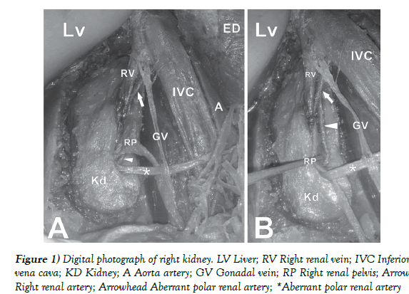
The right kidney had 36.35 mm in width on its anterior face and 52.16 mm in width on its posterior face with 141.22 mm in length. The inferior polo of the right kidney was in contact with the superior margin of the right iliac crest. The main renal artery had originated in the abdominal part of the aorta artery and followed a course of 81.66 mm until its entrance on the renal hilum, with 6.54 mm in size, however instead of a rectilinear trajectory from the aorta it had a descending and quite angular trajectory. From the main renal artery arises an anomalous polar artery that follows a trajectory of 55.59 mm from its origin until its entrance in the inferior polo of the right kidney. This anomalous artery has 2.65 mm in size and starts 51.88 mm after the origin of the main renal artery (Figure 1).
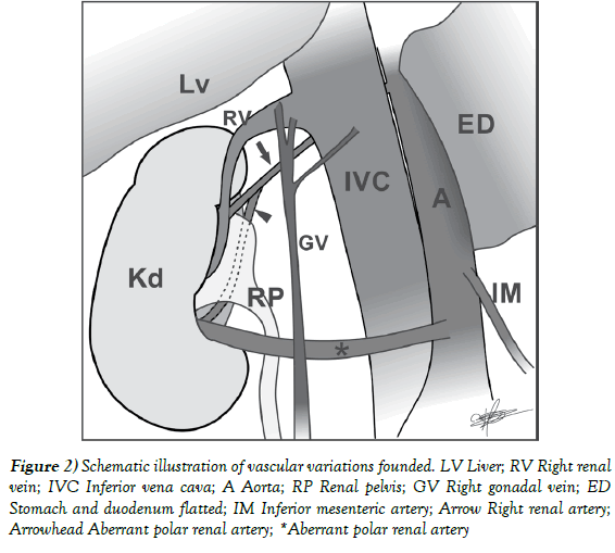
On a lower level, at the same height as the aorta bifurcation arise another anomalous polar artery with 4.93 mm in size and 75.29 mm in length from its origin until its entrance in the renal hilum following a rectilinear trajectory until the inferior polo of the right kidney (Figure 2).
Figure 2: Schematic illustration of vascular variations founded. LV Liver; RV Right renal vein; IVC Inferior vena cava; A Aorta; RP Renal pelvis; GV Right gonadal vein; ED Stomach and duodenum flatted; IM Inferior mesenteric artery; Arrow Right renal artery; Arrowhead Aberrant polar renal artery; *Aberrant polar renal artery
Discussion
As the kidneys ascend from the pelvis, they receive their blood supply from vessels that are close to them, so they are initially vascularized by branches of the common iliac arteries, and then by branches of the distal aorta, and when they finally reach their highest level receive the branches known as renal arteries. In this process of modification in the blood supply from embryonic to fetal life supernumerary branches may arise from the persistence of these primary branches which should involute as the kidneys ascend [4,8].
Several authors already determined anatomical variations from the renal arteries. Budhiraja et al. [4] found multiple pre-hilar branches in 11.66% of its sample, duplication of the renal artery in 8,33% and superior polar arteries in 6,66% of the cases. Glodny et al. [9] in his study through multidetector TC 64-slice determined that 262 patients (24.4%) had 287 additional arteries and 6 accessories on the right side and in a universe of 271 patients he found 298 additional arteries and 6 accessories on the left side. Thakuria et al. [7] found in the same cadaver 4 variant arteries on the right side and 5 variant arteries on the left side, all of them originating from the aorta. Rad [2] in a case study assessed a right anomalous polar artery that entered the anterior face of the kidney towards the superior polo.
Rad [2] suggests that the probability of the presence of additional renal arteries is bigger when the diameter of the main renal artery is lesser than 4.15 mm, being that to the author the renal artery is normally bigger than 5.5 mm without the presence of additional branches. In our specimen the renal artery had 4.15 mm and even so we found an additional branch that followed until the inferior polo of the right kidney.
Budhiraja et al. [1] found a similar variation to ours with an anomalous renal artery originating in the main renal artery and entering the inferior polo of the kidney and called it extra-hilar inferior polar artery, as well as a branch originated in the aorta and entering the inferior polo of the kidney in 4 of 42 studied specimen.
Patasi and Boozary [5] described a similar case to our concerning an accessory renal artery, originating below the superior mesenteric artery in the aorta and following directly to the inferior polo of the right kidney.
We can notice that there is a large amount and variety of accessory and anomalous renal arteries, with different measures and trajectories. There is a vast range of branches that supply the superior and inferior polo of the kidneys originating directly from the aorta or from the main renal artery and that is widely described in literature, however there is no general agreement between the different authors about the nomenclature used in the description of each case.
In order to avoid serious surgical injuries the knowledge of the anatomical uniqueness of each individual becomes indispensable. Surgical planning in renal donors requires careful evaluation of their anatomy, and this study goes beyond transplant surgeries, including aortic artery surgeries, aneurysms, urologic repairs, and angiographic interventions
This detailed map should be constructed using the most modern imaging methods, such as digital subtraction angiography, angiotomography and angioresonance, thus obtaining all the pertinent details that will guarantee the safest intervention, eliminating unnecessary risks to the patient and to all professionals involved.
Conflict of Interest Statement
All authors warrant that there will be no financial and personal relationships with other persons or organizations that may unduly (bias) influence this work.
References
- Budhiraja V, Rastogi R, Jain V, et al. Anatomical variations of renal artery and its clinical correlations: a cadaveric study from central India. J Morphol Sci. 2013;30:228-33.
- Rad T. Aberrant renal arteries and its clinical significance: a case report. Int J Anatom Var. 2011;4:37-9.
- Budhiraja V, Rastogi R, Asthana AK. Variant origin of superior polar artery and unusual hilar branching pattern of renal artery with clinical correlation. Folia Morphol (Warsz). 2011;70:24-8.
- Budhiraja V, Rastogi R, Asthana AK. Renal artery variations: embryological basis and surgical correlation. Romanian J Morphol Embryol. 2010;51:533-6.
- Patasi B, Boozary A. A case report: accessory right renal artery. Int J Anatom Var. 2009;2:155-7.
- Ozkan U, Oğuzkurt L, Tercan F, et al. Renal artery origins and variations: angiographic evaluation of 855 consecutive patients. Diagn Interv Radiol. 2006;12:183-6.
- Thakuria S, Roy RD, Baruah P, et al. Multiple renal arteries: a case report. Int J Anatom Var. 2013;6:155-7.
- Moore KL, Persaud TVN, Torchia, MG. The Developing Human: Clinically Oriented Embryology. Philadelphia: Elsevier, 2015:249.
- Glodny B, Rapf K, Unterholzner V, et al. Accessory or additional renal arteries show no relevant effects on the width of the upper urinary tract: a 64-slice multidetector CT study in 1072 patients with 2132 kidneys. Br J Radiol. 2011;84:145-52.
Ruiz CR, Sergio RRN*, Vidsiunas AK, Souza CC, Lilian A
- *Corresponding Author:
- Sergio RR
São Camilo University Center
São Paulo–SP, Brazil.
Telephone 55-11-2215-2361
E-mail: srrnascimento@gmail.com
Published Online: 2 June 2017
Citation : Ruiz CR, Sergio RRN, Vidsiunas AK, et al. Two aberrant polar arteries to right kidney: A case report. Int J Anat Var. 2017;10(2):016-17.
© This open-access article is distributed under the terms of the Creative Commons Attribution Non-Commercial License (CC BY-NC) (http:// creativecommons.org/licenses/by-nc/4.0/), which permits reuse, distribution and reproduction of the article, provided that the original work is properly cited and the reuse is restricted to noncommercial purposes. For commercial reuse, contact reprints@pulsus.com
Abstract
This study intends to describe an anatomical variation found in a routine dissection of a male cadaver. The main renal artery had originated in the abdominal part of the aorta artery and followed his course until its entrance on the renal hilum; however instead of a rectilinear trajectory from the aorta it had a descending and quite angular trajectory. From the main renal artery arises an anomalous polar artery that enters in the inferior polo of the right kidney. This anomalous artery starts 51,88 mm after the origin of the main renal artery. On a lower level, at the same height as the aorta bifurcation arise another anomalous polar artery that enters in the renal hilum following a rectilinear trajectory until the inferior polo of the right kidney. There is a vast range of branches that supply the superior and inferior polo of the kidneys originating directly from the aorta or from the main renal artery and that is widely described in literature, however there is no general agreement between the different authors about the nomenclature used in the description of each case.
-Keywords
Anatomical variations, Renal artery, Kidney, Vascular variation, Abdomen
Introduction
The renal arteries, arising from the abdominal part of the aorta have variations in number, origin, ramification and trajectory, being that these variations aren’t rare. According to Budhiraja et al. [1] the standard classic formation of an artery and a vein from each side of the individual appears in less than 25% of the cases. Despite the high frequency of variations, there is a large amount of anatomical differences in each variation, making its documentation of extreme importance for the surgical clinic, for the diagnostic imaging beyond the academic field during the training of new professionals [2-6].
There are accessory arteries that follow towards the kidney through the hilum and anomalous arteries that supply the kidney without going through the hilum [2]. These arteries can be directed towards the superior polo or to the inferior polo being in these cases called polar arteries [1,2,7].
Herein we present two aberrant polar arteries to the right kidney originating from the right renal artery and aorta; the left kidney had normal vascular anatomy.
Case Report
On a routine dissection in the Anatomy Laboratory of the São Camilo University Center, with the intention of removing some internal organs from a male cadaver of 65 years old, variations in the quantity of renal arteries were noticed, as well as in their origin being that the right kidney showed visible difference in its shape, size and location compared to the left kidney. The medical history of this cadaver was not available, and the anatomy of left kidney and his vascularization was normal.
The right kidney had 36.35 mm in width on its anterior face and 52.16 mm in width on its posterior face with 141.22 mm in length. The inferior polo of the right kidney was in contact with the superior margin of the right iliac crest. The main renal artery had originated in the abdominal part of the aorta artery and followed a course of 81.66 mm until its entrance on the renal hilum, with 6.54 mm in size, however instead of a rectilinear trajectory from the aorta it had a descending and quite angular trajectory. From the main renal artery arises an anomalous polar artery that follows a trajectory of 55.59 mm from its origin until its entrance in the inferior polo of the right kidney. This anomalous artery has 2.65 mm in size and starts 51.88 mm after the origin of the main renal artery (Figure 1).
On a lower level, at the same height as the aorta bifurcation arise another anomalous polar artery with 4.93 mm in size and 75.29 mm in length from its origin until its entrance in the renal hilum following a rectilinear trajectory until the inferior polo of the right kidney (Figure 2).
Figure 2: Schematic illustration of vascular variations founded. LV Liver; RV Right renal vein; IVC Inferior vena cava; A Aorta; RP Renal pelvis; GV Right gonadal vein; ED Stomach and duodenum flatted; IM Inferior mesenteric artery; Arrow Right renal artery; Arrowhead Aberrant polar renal artery; *Aberrant polar renal artery
Discussion
As the kidneys ascend from the pelvis, they receive their blood supply from vessels that are close to them, so they are initially vascularized by branches of the common iliac arteries, and then by branches of the distal aorta, and when they finally reach their highest level receive the branches known as renal arteries. In this process of modification in the blood supply from embryonic to fetal life supernumerary branches may arise from the persistence of these primary branches which should involute as the kidneys ascend [4,8].
Several authors already determined anatomical variations from the renal arteries. Budhiraja et al. [4] found multiple pre-hilar branches in 11.66% of its sample, duplication of the renal artery in 8,33% and superior polar arteries in 6,66% of the cases. Glodny et al. [9] in his study through multidetector TC 64-slice determined that 262 patients (24.4%) had 287 additional arteries and 6 accessories on the right side and in a universe of 271 patients he found 298 additional arteries and 6 accessories on the left side. Thakuria et al. [7] found in the same cadaver 4 variant arteries on the right side and 5 variant arteries on the left side, all of them originating from the aorta. Rad [2] in a case study assessed a right anomalous polar artery that entered the anterior face of the kidney towards the superior polo.
Rad [2] suggests that the probability of the presence of additional renal arteries is bigger when the diameter of the main renal artery is lesser than 4.15 mm, being that to the author the renal artery is normally bigger than 5.5 mm without the presence of additional branches. In our specimen the renal artery had 4.15 mm and even so we found an additional branch that followed until the inferior polo of the right kidney.
Budhiraja et al. [1] found a similar variation to ours with an anomalous renal artery originating in the main renal artery and entering the inferior polo of the kidney and called it extra-hilar inferior polar artery, as well as a branch originated in the aorta and entering the inferior polo of the kidney in 4 of 42 studied specimen.
Patasi and Boozary [5] described a similar case to our concerning an accessory renal artery, originating below the superior mesenteric artery in the aorta and following directly to the inferior polo of the right kidney.
We can notice that there is a large amount and variety of accessory and anomalous renal arteries, with different measures and trajectories. There is a vast range of branches that supply the superior and inferior polo of the kidneys originating directly from the aorta or from the main renal artery and that is widely described in literature, however there is no general agreement between the different authors about the nomenclature used in the description of each case.
In order to avoid serious surgical injuries the knowledge of the anatomical uniqueness of each individual becomes indispensable. Surgical planning in renal donors requires careful evaluation of their anatomy, and this study goes beyond transplant surgeries, including aortic artery surgeries, aneurysms, urologic repairs, and angiographic interventions
This detailed map should be constructed using the most modern imaging methods, such as digital subtraction angiography, angiotomography and angioresonance, thus obtaining all the pertinent details that will guarantee the safest intervention, eliminating unnecessary risks to the patient and to all professionals involved.
Conflict of Interest Statement
All authors warrant that there will be no financial and personal relationships with other persons or organizations that may unduly (bias) influence this work.
References
- Budhiraja V, Rastogi R, Jain V, et al. Anatomical variations of renal artery and its clinical correlations: a cadaveric study from central India. J Morphol Sci. 2013;30:228-33.
- Rad T. Aberrant renal arteries and its clinical significance: a case report. Int J Anatom Var. 2011;4:37-9.
- Budhiraja V, Rastogi R, Asthana AK. Variant origin of superior polar artery and unusual hilar branching pattern of renal artery with clinical correlation. Folia Morphol (Warsz). 2011;70:24-8.
- Budhiraja V, Rastogi R, Asthana AK. Renal artery variations: embryological basis and surgical correlation. Romanian J Morphol Embryol. 2010;51:533-6.
- Patasi B, Boozary A. A case report: accessory right renal artery. Int J Anatom Var. 2009;2:155-7.
- Ozkan U, Oğuzkurt L, Tercan F, et al. Renal artery origins and variations: angiographic evaluation of 855 consecutive patients. Diagn Interv Radiol. 2006;12:183-6.
- Thakuria S, Roy RD, Baruah P, et al. Multiple renal arteries: a case report. Int J Anatom Var. 2013;6:155-7.
- Moore KL, Persaud TVN, Torchia, MG. The Developing Human: Clinically Oriented Embryology. Philadelphia: Elsevier, 2015:249.
- Glodny B, Rapf K, Unterholzner V, et al. Accessory or additional renal arteries show no relevant effects on the width of the upper urinary tract: a 64-slice multidetector CT study in 1072 patients with 2132 kidneys. Br J Radiol. 2011;84:145-52.


