Unilateral occurrence of extensor medii proprius: an incidental finding
Received: 25-Nov-2020 Accepted Date: Jan 28, 2021; Published: 04-Feb-2021, DOI: 10.37532/1308-4038.14(1).157-158
This open-access article is distributed under the terms of the Creative Commons Attribution Non-Commercial License (CC BY-NC) (http://creativecommons.org/licenses/by-nc/4.0/), which permits reuse, distribution and reproduction of the article, provided that the original work is properly cited and the reuse is restricted to noncommercial purposes. For commercial reuse, contact reprints@pulsus.com
Abstract
The deep extensors muscle of forearm may contain inconsistent muscles like extensor digitorum brevis manus (EDBM), extensor medii proprius (EMP), extensor indicis et medii communis (EIMC) and anomalous extensor indicis proprius (aEIM). We report a rare case of EMP with its unusual course in insertion along with co-existence of extensor indicis proprius muscle (EIP). It originated along with EIP from the distal third of dorsum of ulna and adjacent interosseous membrane, just distal to extensor pollicis longus (EPL). Distally the tendon of the muscle merged with extensor expansion at metacarpophalangeal joint of third digit deep to the extensor digitorum tendon. The knowledge of such variations on the hand is essential to the clinicians and surgeons as hand injuries are one of the most common injuries occurring due to its superficial location and poor insulation. Additionally, this information may help them in accurate diagnosis and management of functional deformities, pathological lesions and various other disorders of hand.
Keywords
Extensor medii; Proprius; Forearm; Extensor expansion; Hand
Introduction
Variations related to muscle of the extensor compartment of forearm are not uncommon and found incidentally during anatomical dissections and surgeries of hand [1,2]. The EIP, a deep extensors muscle of forearm originates from the posterior surface of distal two third of the shaft of ulna and adjacent interosseous membrane. The muscle tendon passes inferiorly and inserts at the extensor expansion of proximal phalanx of the second digit ulnar to the tendon of extensor digitorum [3]. This muscle gives off several accessory slips to the extensor tendons of other fingers and may contain anomalous muscles like extensor digitorumbrevismanus (EDBM), extensor mediiproprius (EMP), extensor indicis et mediicommunis (EIMC) and anomalous extensor indicisproprius (aEIM) [4]. The aEMP is an analogous muscle to the EIP being inserted to the dorsal expansion of third digit instead of the second digit. It often co-exists with the EIP muscle or with its variant and has an incidence rate between 0.8% to 12%. Being asymptomatic, its presence may mislead the clinicians and surgeons during diagnosis of hand diseases and treatment [5,6].
We present a case of EMP with its unusual course while inserting to the extensor expansion of the middle finger along with co-existing of aEIM.
Case Report
During a routine anatomical dissection of upper limb, an unusual muscle in the right hand of a 60-yr-old male cadaver was identified. A midline incision was made on the dorsum of the forearm and hand, from the lower one third of the arm to the distal phalanx of middle finger. The skin along with superficial fascia was reflected and related neurovascular structures were exposed. The thickened deep fascia on the dorsum of the wrist along with its modification, the extensor retinaculum was noted. The margins and attachments of the retinaculum forming the osseofascial compartments were thoroughly observed. Six osseofascial compartments were formed by the septa from retinaculum for the passage of extensor tendons from the forearm to digits.
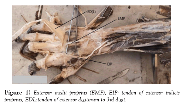
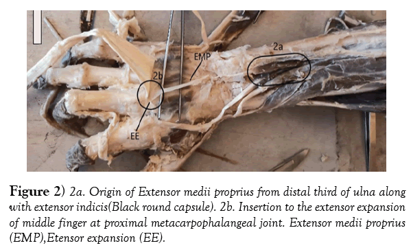
In order to further observe the underlying superficial and deep muscles of forearm a longitudinal incision was made on the retinaculum followed by reflecting the extensor digitorum (ED) tendon distally. We observed an additional muscle tendon inserting to extensor expansion of middle finger. Its muscle fibres were arising along with extensor indicis (EI) from the distal third of dorsum of ulna and adjacent interosseous membrane, just distal to extensor pollicislongus. The length of the muscle belly and tendon was 3.6 cm and 9.2 cm respectively. The long slender tendon coursed downwards from the myotendinous junction in the fourth extensor compartment along with the ED and EI tendons. No additional tendon slips to the middle finger was seen. The insertion of this variant muscle was observed on the extensor expansion at metacarpophalangeal joint of third digit, deep to the extensor digitorum tendon. We could not trace the nerve supply to the EMP. (Figures 1 & 2) shows the origin and insertion of the variant muscle with the presence of EIP muscle. The dissected limb region of right forearm did not show any other variations, trauma nor surgical procedures on it.
Discussion
A review of literature shows that the described muscle is an aberrant muscle and analogues to the extensor indicis, likely found mostly in cadaveric dissections, a plane deep to the extensor digitorum tendons. This anomaly termed as the EMP, also known as the extensor mediidigiti, with two forms: longus and a brevis. The muscle described in our case is most likely to belong on the basis of insertion made to the extensor expansion of the middle finger [7,8]. It often co-exists with the extensor indicis muscle or with its variant. Von Schroeder HP and Botte MJ, reported that the EMP and extensor indicisproprius muscles had a common origin with separable muscle belly in all the fifty-eight adult hands they studied, which is similar to our case report [9].
The EMP has an incidence ranging between 1% and 12% in cadaveric studies and observed more frequently in males than females. A meta-analysis by Kaissar Yammine revealed that Indian populations showed lowest rate of EMP compared to Japanese, Europeans and North Americans [2,6,9]. In the present case, The EMP originates from the posterior surface of distal one third of Ulna with adjacent interosseous membrane and ulnar to the origin of the extensor indicis. Some authors reported that, the EMP also arises from the distal part of muscle belly of Extensor indices [10], Holmes et al. observed the course of EMP tendon while passing deep to the intertendinous connections of the extensor digitorum muscle, arising from the lunate bone, whereas in our case it was seen deep to the extensor digitorum tendon [11]. The frequent insertion of the tendon is reported on the dorsal expansion of the middle finger, usually on the palmer and ulnar aspects of the Extensor digitorum tendon. While in the present case, tendon was palmar to the extensor digitorum tendon, on inserting to the extensor expansion of metacarpophalangeal joint of middle finger. This type of insertion is less reported in literature and rarely found [1,5]. In this case report, we could not identify the nerve supply of the EMP. However, Studies available in literature is shown that the EMP was innervated by the branches of posterior interosseous nerve [8,11].
Embryological explanation
The upper limb muscles develop from mesoderm derived from myotomes, after migrating into the limb bud during the 5th week of intrauterine life. This mesoderm differentiates into anterior and posterior condensations. The extensor and supinator muscles of the upper limb are derived from the posterior condensations [12]. However, the posterior condensations further differentiate into three parts and forms superficial, radial potion and deep layers. The deep layer of the extensor musculature shows marked phylogenetic instability and undergoes variations. Based on phylogenetic comparisons, the origin of extensor medii proprius muscle may be an evolutionary remnant of a normal developmental arrangement [13,14]. Functionally, the passive traction of the EMP tendon resulted in extension of middle finger and may add additional strength to Extensor digitorum tendon during extension of metacarpophalangeal joint. As reported by Holmes et al, the active traction of EMP may have negligible effect on the metacarpophalangeal extension due to small sized muscle belly with thin tendon [11].
Conclusion
Muscular variations of the extensor compartment of forearm are uncommon and may result in restricted movements of forearm and hand. The EMP is one of the aberrant muscles of forearm, usually asymptomatic and reported incidentally during the anatomical dissections and in hand surgeries. The reported muscle in our case had rare variety of insertion, which is less documented in literature. The knowledge of such variations of the hand is essential to the clinicians and surgeons in diagnosis of hand pathologies and surgeries related to tendon transfer or repair.
Acknowledgement
The authors wish to express their sincere gratitude to Principal and the management committee of Grameen Ayurvedic Medical College, Terdal, Karnataka state, India for providing necessary facilities to carry out this work.
REFERENCES
- Tan ST, Smith PJ. Anomalous extensor muscles of the hand: a review. J Hand Surg Am. 1999;24:449-55.
- Yammine K. The prevalence of the extensor indicis tendon and its variants: a systematic review and meta-analysis. Surg Radiol Anat. 2015;37:247-54.
- Standring S. Gray’s anatomy. The anatomical basis of clinical practice. 41st ed. New York, Elsevier. 2016;855.
- Komiyama M, New TM, Toyota N, et al. Variations of the extensor indicis muscle and tendon. J Hand Surg Br. 1999;24:575-8.
- Klena JC, Riehl JT, Beck JD. Anomalous extensor tendons to the long finger: a cadaveric study of incidence. J Hand Surg Am. 2015;37:938-41.
- https://en.wikipedia.org/wiki/Extensor_medii_proprius_muscle
- Von Schroeder HP, Botte MJ. Anatomy of the extensor tendons of the fingers: variations and multiplicity. J Hand Surg Am. 1995;20:27-34.
- Carlos JS, Goubran E, Ayad S. The presence of extensor digitimedii muscle-anatomical variant. J Chiropr Med. 2011;10:100-4.
- Von Schroeder HP, Botte MJ. Anatomy of the extensor tendons of the fingers: variations and multiplicity. J Hand Surg Am. 1995;20:271141-5.
- Stanchev S, Iliev A, Malinova L, et al. A Rare Case of Unusual Origin of Extensor Medii Proprius Muscle and its Clinical Significance. Actamorphologica et anthropologica. 2017;24:3-4.
- Liu HH, Rosales A, Kirchhoff CA. A Variant of Extensor Medii Proprius: A Case Report. 2015.
- SinghV. Textbook of Clinical Embryology. New Delhi, Elsevier. 2012;103-107.
- Souter W. The extensor digitorumbrevismanus. Br J Surg. 1966;53:821-3.
- Meenakshi Khullar SS, Morphology of Extensor Indicis Proprius Muscle in the North Indian Region: An Anatomic Study. Int J Anat Radiol Surg. 2019;8:AO17-AO21.


