Unknown foramen found in the middle cranial fossa
2 Department of Basic Sciences, University of Rio dos Sinos Valley, Sao Leopoldo, Rio Grande do Sul, Brazil
3 Department of Basic Sciences, University of Santa Cruz do Sul, Brazil
4 Department of Basic Sciences, Federal University of Sergipe, Brazil
5 Department of Basic Sciences, Tiradentes University, Brazil
Received: 16-Oct-2019 Accepted Date: Feb 18, 2020; Published: 25-Feb-2020, DOI: 10.37532/1308-4038.20.13.82
Citation: Bonatto-Costa JA, Campos DD, Aragao JA, et al. Unknown foramen found in the middle cranial fossa. Int J Anat Var. 2020;13(1): 82-83.
This open-access article is distributed under the terms of the Creative Commons Attribution Non-Commercial License (CC BY-NC) (http://creativecommons.org/licenses/by-nc/4.0/), which permits reuse, distribution and reproduction of the article, provided that the original work is properly cited and the reuse is restricted to noncommercial purposes. For commercial reuse, contact reprints@pulsus.com
Abstract
The sphenoid bone is located in the middle cranial fossa and has a lot of structures going through its foramina. However, in a skull belonging to the collection of the anatomic laboratory of the University of Rio dos Sinos Valley, an unusual foramen was found. It was placed below the optic canal, in the body of the sphenoid bone, thus connecting the middle cranial fossa with the nasal cavity and it was not yet described in the literature. Observing this, it was decided that the next step should be a search in the collection of another two universities in the Brazilian South and one in the Northeast. The presence of the unknown foramen was checked in dry skulls with their base exposed, verifying its prevalence in the collection of the anatomic labs of the participating universities. Following the inclusion criteria, another 96 skulls were analyzed, looking for the same observed foramen, and was found in another 4 skulls, making a total of 5.15%. Among them, two had presented it unilaterally and the other three, bilaterally. Because the analysis was performed in dry skulls, it was not possible to observe probable structures that could be going through it. Knowing its existence will benefit various researchers and other professionals of the field. Future perspectives for this study will be based on the observation of this foramen in new cadavers donated to these institutions during the dissection and removal of brains to try to see what structures may be passing by it.
Keywords
Sphenoid bone; Nasal cavity; Skull; Middle cranial fossa; Pituitary gland
Introduction
The knowledge of the positions of many orifices and fissures found in the sphenoid bone is of the utmost importance in the clinical and surgical practice, because they accommodate a lot of blood vessels and nerves [1,2]. It is also known that a broad spectrum of congenital abnormalities affects the cranial base, one of them, the persistent pituitary canal, rare congenital defect that allows the pituitary gland to present itself as a nasopharyngeal [3,4]. Various reports have been described about the variation of different foramina in the skull, however, until now little to no reports have been described about a foramen that communicates the middle cranial fossa with the nasal cavity. Besides that, there was no embryonic malformation describing what was seen [5,6]. Therefore, in a succinct description using as a base the existent literature–of the area where the variation was found, it is noticed that the foramen observed is indeed atypical. So, the main goal of this work was to determine the presence of the said foramen in dry human skulls.
Case Report
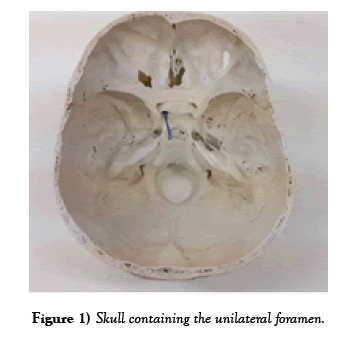
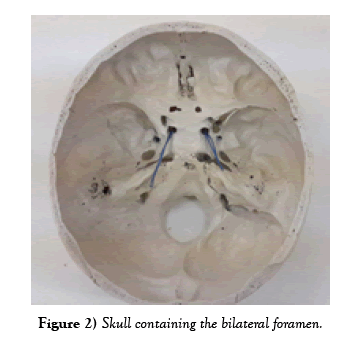
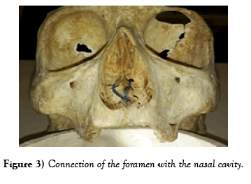
It was realized in a survey of 97 dry human skulls, in three universities of the Brazilian South and one of the Northeast, concerning an variating foramen that communicate the middle cranial fossa with the nasal cavity. Of the 97 skulls examined (Table 1), it was found the uncommon foramen in 5, 15% [5]. It was presented both uni and bilaterally (Figures 1 & 2), being, most of the times, in circular shape, located in the body of the sphenoid bone, anterolateral to the sella turcica and inferior to the optic canal (Figure 3).
| University | N | Unilateral foramen | Bilateral foramen |
|---|---|---|---|
| UFCSPA | 22 | 0 | 0 |
| UNISC | 28 | 0 | 0 |
| UNISINOS | 22 | 2 | 2 |
| UFS | 25 | 0 | 0 |
| TOTAL | 97 | 2 | 3 |
Table 1 Frequency of the unusual foramen in the cranial base.
Discussion
These variations of the base of the skull foramina are very common, involving duplicated foramina, abnormal openings, asymmetry, canalization and absence. Those usually cause a lot of confusion during surgical intervention in the region [7-9].
Several authors have reported variations in the morphology of the foramina in the base of the skull, like atypical foramen ovale, a rare defect in the craniopharyngeal canal, well corticated of the sphenoid body in the medium line of the sella turcica floor to the nasopharynx roof [1]. Although the embryogenesis of the canal has been contested, evidence supports its origin from the incomplete closing of the Rathke’s pouch, the precursor of the adenohypophysis [3,10,11].
In this word, it was observed an unusual foramen of smooth and circular outline in five (5,15%) adult skulls of two Brazilian regions that communicated the middle cranial fossa with the nasal cavity [12]. A foramen with the same characteristics was also reported for the first time by Nayak in an isolated cranial sample; however he did not mention a communication with the nasal cavity. Comprehending the basic anatomy and abnormal variation of the cranial base will not allow interventional radiologists and surgeons to make mistakes, being on imaging interpretation or cranial surgeries. Which implicates safety, efficiency and comprehensive knowledge of anatomy, morphological variations and morphometric values of the cranial base structures [13].
Conclusion
Being the analysis made in dry skulls and having no previous record of the information in the literature, no knowledge about possible structures passing through this foramen was obtained. The presence of the foramen in the other analyzed skulls, including those from a different region of the country, suggests that this anatomical anomaly can present some function or may be related to ethnicity or can also be a genetic condition. A study showing a forensic analysis of this skulls and the determination of ethnicity and sex could help investigate a possible reason for the appearance of this foramen. The knowledge of its existence shows that there is need for further research. Connecting the presence of the foramen with a pattern on its emergence could help understand how it emerged and for what purpose. It would be also important of identify possible structures going through it as well as its functions. For this, as future perspectives, it would be ideal a survey in corpses donated to the participating institutions during its first contact with dissection, in order to try identifying the foramen with the structure passing through it as its functions.
Acknowledgments
The authors express heartfelt gratitude to all the academics, professors and lab technicians of the Department of Anatomy of their respective universities. The paper has not been submitted for publication elsewhere and all authors were fully involved in the study and preparation of the manuscript. There is not conflict of interest also.
REFERENCES
- Skrzat J, Walocha J, Srodek R, et al. An atypical position of the foramen ovale. Folia Morphol (Warsz). 2006;65:396-9.
- Abd Latiff A, Das S, Sulaiman IM, et al. The accessory foramen ovale of the skull: an osteological study. Clin Ter. 2009;160:291-3.
- Hughes ML, Carty AT, White FE. Persistent hypophyseal (craniopharyngeal) canal. Br J Radiol. 1999;72:204-6.
- Abele TA, Salzman KL, Harnsberger HR. Craniopharyngeal canal and its spectrum of pathology. Am J Neuroradiol. 2014;35:772-7.
- SADLER, Thomas W. Langman embriologia médica. 12. ed. Rio de Janeiro: Guanabara Koogan, 2013.
- MOORE, Keith L. Anatomia orientada para a clínica. 7. ed. Rio de Janeiro: Guanabara Koogan, 2014.
- Reymond J, Charuta A, Wysocki J. The morphology and morphometry of the foramina of the greater wing of the human sphenoid bone. Folia Morphol (Warsz). 2005;64:188-93.
- Shinohara AL, de Souza Melo CG, Silveira EM, et al. Incidence, morphology and morphometry of the foramen of Vesalius: complementary study for a safer planning and execution of the trigeminal rhizotomy technique. Surg Radiol Anat. 2010;32:159-64.
- Mazengenya P, Ekpo O. Unusual foramen in the middle cranial fossae of adult black South African skull specimens. Surg Radiol Anat. 2017;39:815-8.
- Hori A, Schmidt D, Rickels E. Pharyngeal pituitary: development, malformation, and tumorigenesis. Acta Neuropathol. 1999;98:262-72.
- Kaushik C, Ramakrishnaiah R, Angtuaco EJ. Ectopic pituitary adenoma in persistent craniopharyngeal canal: case report and literature review. J Comput Assist Tomogr. 2010;34:612-4.
- Nayak S. An abnormal foramen connecting the middle cranial fossa with sphenoidal air sinus: A Case Report. J Biol Anthropol 2007;2:1-3.
- Keskil S, Gozil R, Calguner E. Common surgical pitfalls in the skull. Surg Neurol. 2003;59:228-31.


