Unusual bilateral variation in the origin, number and distribution pattern of pectoral nerves
Received: 23-Mar-2021 Accepted Date: Apr 12, 2021; Published: 19-Apr-2021, DOI: 10.37532/1308-4038.14(4).193-195
Citation: Tessema CB. Unusual Bilateral Variation in the Origin, Number and Distribution Pattern of Pectoral Nerves. Int J Anat Var. 2021;14(4):79-81.
This open-access article is distributed under the terms of the Creative Commons Attribution Non-Commercial License (CC BY-NC) (http://creativecommons.org/licenses/by-nc/4.0/), which permits reuse, distribution and reproduction of the article, provided that the original work is properly cited and the reuse is restricted to noncommercial purposes. For commercial reuse, contact reprints@pulsus.com
Abstract
During the dissection of a 69-year-old male cadaver, three pectoral nerves from the trunks and divisions of the brachial plexus were detected bilaterally. Superior pectoral nerves arose from the anterior divisions of the superior trunks. The middle pectoral nerve originated from the middle trunk on the right side and from the anterior division of the middle trunk on the left side and inferior pectoral nerves branched from the inferior trunks. What is peculiar in this case is the bi-laterality and the division of the middle pectoral nerves into anterior and posterior branches, where the posterior branches joined the inferior pectoral nerves to form a short common stem that innervated both pectoralis muscles.
Such variations could predispose the nerves to unintentional but preventable injuries during surgical, anesthesiologic and other clinical procedures, hence, clinicians should be aware of such variations of pectoral nerves to avoid technical errors, unintended injuries and treatment failures.
Keywords
Brachial plexus, Superior pectoral nerve, Middle pectoral nerve, Inferior pectoral nerve, Common stem
Introduction
It is a common textbook description that two pectoral nerves arise from the medial and lateral cords of the brachial plexus as medial and lateral pectoral nerves. The medial pectoral nerve innervates both pectoralis muscles while the lateral pectoral nerve innervates only the clavicular part of pectoralis major muscle. However, there are well-documented wide range of variations in the origin, number and distribution pattern of pectoral nerves.
According to current literature the pectoral nerves originate not only from the two cord of the brachial plexus but they can have variable origins from the trunks, cords and divisions of the brachial plexus or a combination of these [1]. There are also numerous other studies and reports that demonstrated the wide range of variation in the origin of the individual pectoral nerves thatincluded the origin of medial pectoral nerve from the lower trunk of the brachial plexus and the lateral pectoral nerve from the anterior divisions of the superior and middle trunks [2]. There are also several other related documentations of variations in the origin and number of the pectoral nerves like the consistent presence of three (superior, middle and inferior) pectoral nerves arising from the cords and division of the brachial plexus [3], two lateral pectoral nerves arising from the lateral cord of the brachial plexus [4], three separate lateral pectoral nerves arising from the lateral cord of the brachial plexus [5] and lateral pectoral nerve originating from the anterior division of the superior trunk and medial pectoral nerve from the middle trunk of the brachial plexus [6].
The pectoral nerves also vary in their distribution to the pectoralis muscles. In ideal circumstances, the medial pectoral nerve innervates the pectoralis minor and one of its branch’s pierces through the pectoralis minor muscle and innervates the pectoralis major muscle. However, it is known that more branches of the medial pectoral nerve pierce pectoralis minor muscle and others may under cross it to innervate the inferior portion of the sternocostal part and abdominal part of the pectoralis major muscle. The lateral pectoral nerve often sends a small communicating branch to the medial pectoral nerve to form ansa pectoralis through which it contributes to the innervation of pectoralis minor muscle [7,8]. Absence of lateral pectoral nerve with three pectoral nerves arising from the anterior division of the superior trunk and medial pectoral nerve arising from the medial cord with a prominent ansa pectoralis that provided branches to both pectoralis muscles was also recently reported [9].
Due to these variabilities, the pectoral nerves are clinically important in that they can be involved in various diseases, traumatic and entrapment injuries of the thorax, shoulder and axilla. They are frequently affected by mononeuropathies and particularly lesions of the lateral pectoralnerve due to repetitive forceful macro- and micro-traumas results in focal muscle atrophy and weakness of the clavicular part of pectoralis major muscle [10].
Case Report
During dissection of the brachial plexus in a 69-year-old male cadaver three nerves to the pectoralis muscles with unusual origin, number and branching patterns were incidentally noticed bilaterally. These nerves were cleaned and followed farther from their origins to their termination on the pectoralis minor and pectoralis major muscles to demonstrate their distribution. Photographs were taken for illustrations and based on their origin and position, the three nerves on each side were designated as superior, middle and inferior pectoral nerves for a descriptive purpose.
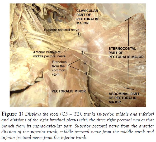
1) Superior pectoral nerves - originated from the anterior divisions of the superior trunks of the right and left brachial plexuses and entered the deep surfaces of the clavicular parts of pectoralis major (Figures 1-5).
Figure 1) Displays the roots (C5 – T1), trunks (superior, middle and inferior) and divisions of the right brachial plexus with the three right pectoral nerves that branch from its supraclavicular part. Superior pectoral nerve from the anterior division of the superior trunk, middle pectoral nerve from the middle trunk and inferior pectoral nerve from the inferior trunk.
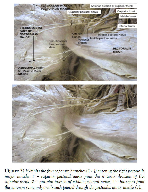
Figure 3) Exhibits the four separate branches (1 - 4) entering the right pectoralis major muscle; 1 = superior pectoral nerve from the anterior division of the superior trunk, 2 = anterior branch of middle pectoral nerve, 3 = branches from the common stem; only one branch pierced through the pectoralis minor muscle (3).
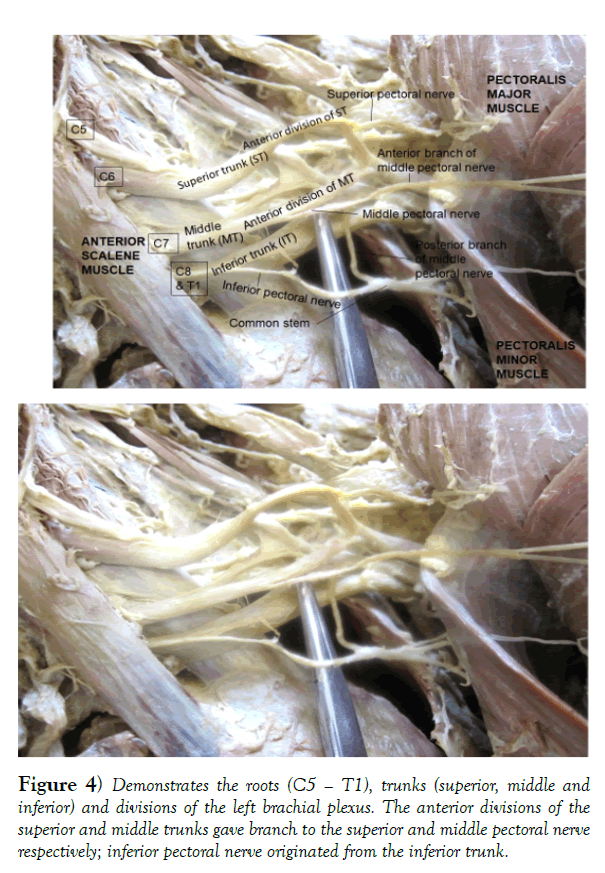
Figure 4) Demonstrates the roots (C5 – T1), trunks (superior, middle and inferior) and divisions of the left brachial plexus. The anterior divisions of the superior and middle trunks gave branch to the superior and middle pectoral nerve respectively; inferior pectoral nerve originated from the inferior trunk.
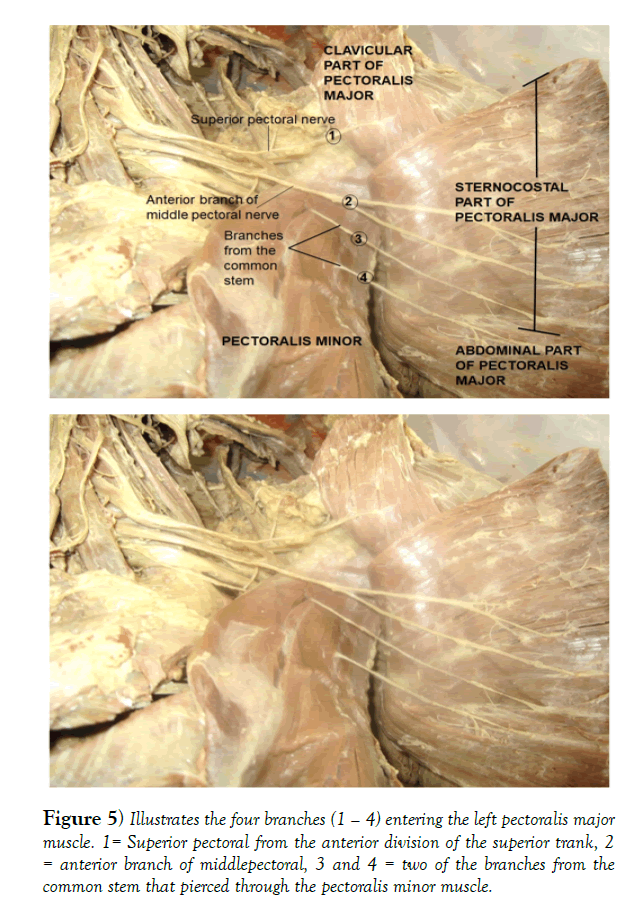
Figure 5) Illustrates the four branches (1 – 4) entering the left pectoralis major muscle. 1= Superior pectoral from the anterior division of the superior trank, 2 = anterior branch of middlepectoral, 3 and 4 = two of the branches from the common stem that pierced through the pectoralis minor muscle.
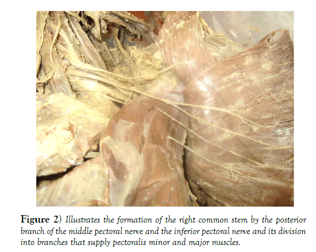
2) Middle pectoral nerves - arose from the middle trunk of the brachial plexus on the right side (Figures 1-3) and from the anterior division of the middle trunk on the left side (Figure 4). At the medial borders of the right and left pectoralis minor muscles, the middle pectoral nerves split into anterior and posterior branches. The anterior branches ran superficial to the proximal parts of pectoralis minor muscles and entered the superior portions of sternocostal parts of pectoralis major muscles and the posterior branches joined the inferior pectoral nerves on both sides to form short common stems that divided and distributed to the two pectoralis muscles (Figures 2 & 4). A remarkable difference is noted in the further course of the branches of the right and left common stems. On the right side the common stem divided into two major branches before entering the pectoralis minor muscle (Figures 2 & 4). One of the branches provided multiple small branches to the pectoralis minor muscle,one of these small branches pierced through it to extend to the middle portion of the sternocostal part of pectoralis major muscle (Figure 3) and the second branch from the common stem ran inferolaterally deep to pectoralis minor and crossed its lateral border to enter the abdominal part of pectoralis major (Figures 2 & 3). On the left side the common stem divided into multiple branches before entering the pectoralis minor muscle. Many of these branches terminated in the pectoralis minor muscle, while two branches pierced the muscle to enter the middle and inferior portion of the sternocostal and abdominal parts of the pectoralis major muscle (Figures 4 & 5).
3)Inferior pectoral nerves - branched from the inferior trunks of the right and left brachial plexuses and coursed laterally deep to pectoralis minor muscles where they are joined by the posterior branches of the middle pectoral nerves to form the short common stems that divided to supply both pectoralis minor and pectoralis major muscles as described above (Figures 1, 2 & 4).
No distinct formation of ansa pectoralis is detected on both sides.
Discussion
Though variations of origin and distribution of pectoral nerves are well documented by different authors, there are similarities and differences between them regarding their farther courseand distribution. Three pectoral nerves (superior, middle and inferior), which are equivalents of the superior, middle and inferior pectoral nerves in this report, were consistently found in 26 brachial plexuses of 15 cadavers, where the superior pectoral nerve arose from the anterior division of the superior trunk or the lateral cord and entered the clavicular part of pectoralis major. A middle branch that coursed on the under-surface of pectoralis major muscle to innervate its sternal part and an inferior branch passed beneath the pectoralis minor to innervate the pectoralis minor muscle and the costal aspect of the pectoralis major muscle were also reported [3]. The farther course of the superior pectoral nerve in the present case is similar to those reported by these authors, while it is different for the middle and inferior pectoral nerves.
Nerves to the clavicular part of pectoralis major are described by many of the authors as well. According to an earlier documentation the lateral pectoral nerve coursed on the undersurface of pectoralis major muscle, innervating the proximal one-third or more of the muscle. While, in 62% of the patients, the medial pectoral nerve coursed through the pectoralis minor muscle to innervate the lower half to two-thirds of the pectoralis major muscle. In the remaining 38% of patients the medial pectoral nerve exits around the lateral aspect of the pectoralis minor muscle [7]. However, in the current case report combinations of both forms are observed on the right side while two branches pierced through the pectoralis minor muscle on the left side. The lateral pectoral nerve can arise from the anterior divisions of the upper and middle trunks or by a single root from the lateral cord of the brachial plexus and has a constant course along the thoracoacromial vessels [8], which is consistent with the superior pectoral nerve in this case report. In another study, the medial pectoral nerve pierced the pectoralis minor as a single trunk to innervate pectoralis major in 56% of the cases, and in 44% of the case it divided before entering pectoralis minor and its branches passed through the pectoris minor muscle or around its lower border to reach pectoralis major [8]. In a previous case of pectoral nerves in our dissection lab, five nerves to the pectoralis major muscle were reported, four of which were from the ansa pectoralis. Out of the four branches from ansa pectoralis, only one branch pierced the pectoralis minor to reach the sternocostal part of pectoralis major [9]. In this current case, four branches to the pectoralis major were observed out of which three of them originated from a common stem but only one of those branches on the right side and two of them on the left side pierced through the pectoralis minor.
The findings in this current case are very unique and different from the long-stablished description of medial and lateral pectoral nerves from the medial and lateral cords in the infraclavicular part of the brachial plexus and any of the documented variations so far in: 1) being bilateral from the supraclavicular part of the brachial plexus, 2) the division of the middle pectoral nerve into anterior and posterior branches and the direct extension of the anterior branches to superior portion of the sternocostal part pectoralis major and 3) the union of the posterior branches of the middle pectoral nerve with the inferior pectoral nerves to form a common stems that divided further into multiple branches that innervated both pectoralis minor and major muscles.
Conclusion
The observation in this case does not match up to the origin, number and distribution pattern of any of the variations reported as yet. Such unusual variations can be sources of uncertainty during performing invasive diagnostic, therapeutic, cosmetic and other clinical procedure on the thoracic wall predisposing the nerves to unintended injuries. Therefore, equipping surgeons, anesthesiologist, interventional radiologists and other clinicians with the awareness of such variations would help in the diagnosis and decision-making processes and intaking appropriate precautionary measures prior to and during actual clinical procedure to avoid or minimize technical errors, iatrogenic injuries and treatment failures.
Acknowledgements
I would like to thank the donor and his families for their invaluable consent and the department of biomedical sciences for its encouragement and support. I am also grateful to Denelle Kees, Chelsy Swanson and John Opland for their technical assistance during the preparation and dissection of this cadaver.
Disclaimer
The opinions or assertions contained here in are the private ones of the author/speaker and are not to be construed as official or reflecting the views of the Department of Defense, the Uniformed Services University of the Health Sciences or any other agency of the U.S.Government.
REFERENCES
- Porzionato A, Macchi V, Stecco C, et al. Surgical anatomy of pectoral nerves and pectoral musculature. Clin Anat. 2012;25:559-75.
- Prakash KG, Saniya K. Anatomical Study of Pectoral Nerves and its Implications in Surgery. J Clin Diagn Res. 2014;8:AC01-AC05.
- David S, Balaguer T, Baque P, et al. The anatomy of the pectoral nerves and its significance in breast augmentation, axillary dissection and pectoral muscle flaps. J Plast Reconstr Aesthet Surg. 2012;65:1193-8.
- Goel S, Rustagi SM, Kumar A, et al. Multiple unilateral variations in medial and lateral cords of brachial plexus and their Branches. Anat Cell Biol. 2014;47:77-80.
- Rai R, Ranade AV, Prabhu LV, et al. Accessory lateral pectoral nerves supplying the pectoralis major. Rom J Morphol Embryol. 2008;49:577-9.
- Shetty P, Nayak SB, Kumar N, et al. Origin of Medial and Lateral Pectoral Nerves from the Supraclavicular Part of Brachial Plexus and its Clinical Importance – A Case Report. J Clin Diagn Res. 2014;8:133-4.
- Hoffman GW, Elliott LF. The Anatomy of the Pectoral Nerves and its Significance to the General and Plastic Surgeon. Ann Surg. 1987;205:504-6.
- Macchi V, Tiengo C, Porzionato A, et al. Medial and Lateral Pectoral Nerves: Course and Branches. Clin Anat. 2007;20:157-62.
- Tessema CB. Aberrant origin and distribution of pectoral nerves with prominent ansa pectoralis and absent lateral pectoral nerve. Int J Anat. 2020;13:1-3.
- Goncalves AdV, Teixeira LC, Torresan R, et al. Randomized Clinical trial on preservation of medial pectoral nerve following mastectomy due to breast cancer: impact on upper limb rehabilitation. Sao Paulo Med J. 2009;127:117-21.


