Unusual case of a superficial radial artery
Received: 01-Jun-2021 Accepted Date: Jun 21, 2021; Published: 29-Jun-2021, DOI: 10.37532/1308-4038.14(6).136-137
Citation: Nelson Davis, Robert Hage. Unusual case of a superficial radial artery. Int J Anat Var. 2021;14(6):103-105.
This open-access article is distributed under the terms of the Creative Commons Attribution Non-Commercial License (CC BY-NC) (http://creativecommons.org/licenses/by-nc/4.0/), which permits reuse, distribution and reproduction of the article, provided that the original work is properly cited and the reuse is restricted to noncommercial purposes. For commercial reuse, contact reprints@pulsus.com
Abstract
While browsing through a metal storage container with prosected specimens, an upper limb with a rare variant of the course of the radial artery was observed. The radial artery ran superficial to the tendons of the abductor pollicislongus and extensor pollicisbrevis and longus forming the anatomical snuff box, instead of underneath (deep to) the tendons
Keywords
Indicis artery, carpal ligament, femoral catheterization
Introduction
This variation is described 19 times [1-5] but has never been documented by photography. The radial artery gave off a medial branch (at the distal one quarter of the forearm) which passed over the carpal ligament to continue as the lateral palmar digital artery of the thumb. The median artery, branching off the common interosseus artery, accompanied the median nerve in the forearm and joined the radialisindicis artery in the palm of the hand. No variations of the arteries proximal to the elbow joint were observed
The radial artery is generally described as a direct continuation of the brachial artery with its course starting 1 cm distal to the flexion crease at the elbow joint [6-8]. Usually smaller in diameter than the ulnar artery, it travels deep to the brachioradialis muscle in the proximal part of the forearm, medial to the neck of the radius. As it courses distally, it is located anterior to the radius where it can be palpated at the wrist. From here it travels dorsally deep within the ‘anatomical snuff box’ between the two heads of the first dorsal interosseous muscle emerging anteriorly, anastomosing with the deep branch of the ulnar artery medially, forming the deep palmar arch.
Clinically, the radial artery is used as a pathway for cardiac catheterization since the procedure is minimally invasive compared to femoral catheterization. There is a low risk for blood loss and nerve injury since the artery is not accompanied by a major nerve. Due to the artery being superficial, underneath the skin, a compression device placed around the wrist after catherization prevents blood loss and allows the patient to be mobile [3].
Methods and Materials
A right upper limb already partially dissected and used as a prosection specimen showed a superficial radial artery as variation. The limb was from a 69-year-old male donor. Embalming details, according to specifications received from our source for donor bodies, shows ¬that 10 gallons of a solution containing 25% of an 85% phenol solution, 25% of diethylene glycol, 25% water and 25% alcohol, of which 1/3 methanol and 2/3 ethanol was used with the addition of 8 ounces of formalin to each 3 gallons.
Since multiple variations in one extremity can be found the arterial branching pattern of the shoulder and upper arm was scrutinized and no variations were observed. Further dissection of the radial artery in the hand was undertaken and documented by taking photographs with a Nikon D7100. Research on cadaveric material is exempted by the St. George’s University IRB under document no 06014. Arteries of the forearm and palm were fully dissected exposing the median artery, palmar arches and digital vessels. The external diameter of the arteries at various locations in the forearm and hand were noted by using a Fowler 6” Vernier Caliper with fine adjustment to one decimal place. Several measurements were taken at specific points along the artery, using the external diameter and little adventitia removed, with the averages recorded in (Table 1).
| Artery | Location | Diameter (mm) |
|---|---|---|
| Radial | cubital fossa | 4.4 |
| middle of forearm | 3.5 | |
| first inter-metacarpal space | 3 | |
| Superficial palmar branch of radial | bifurcation proximal to radial head | 1.7 |
| wrist before it crosses the extensor tendons of the thumb | 1.5 | |
| before anastomosis in the palm (distal to adductor pollicis) | 1.7 | |
| Deep palmar branch of radial | after crossing oblique head of adductor pollicis | 2.3 |
| Median | bifurcation at the cubital fossa | 1.7 |
| middle of forearm | 1.3 | |
| Ulnar | cubital fossa (smaller branch from common interosseous artery) | 2.4 |
| middle of forearm | 1.8 | |
| palm, distal to Guyon’s canal | ||
| Deep branch of ulnar | after crossing flexor digiti minimi and adductor digiti minimi | 1.2 |
Table 1 Diameters of radial artery, its branches, and median and ulnar arteries.
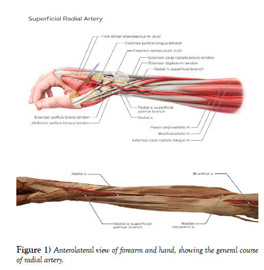
Figure 1 shows the dissected forearm with the brachial artery branching into the radial distal to the bicipitalaponeurosis. The radial artery (diameter 3.5 mm) under cover of the brachioradialis muscle gives of a medial branch (diameter 1.7 mm) in the distal quarter of the forearm 7.3 cm proximal to the styloid process of the radius. The radial artery continues (diameter 3 mm) and crosses over the tendons of the abductor pollicislongus, extensor pollicisbrevis and extensor pollicislongus (diameter 3 mm) and travels between the two heads of the first dorsal interosseus muscle. Here it splits into two branches, the princepspollicis traveling laterally and the radialisindicis medially (diameters 1.9 mm and 2.5 mm respectively).
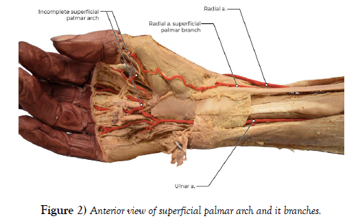
The medial branch from the radial artery (superficial palmar branch) continues distally and passes over the carpal ligament anteriorly into the palm of the hand and provides muscular branches to the thenar region and continues as the lateral palmar digital artery of the thumb (Figure 2).
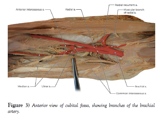
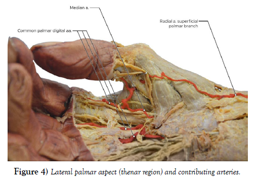
The common interosseous artery, at the cubital fossa, gave rise to a median artery (diameter 1 mm) accompanying the median nerve, then traveling through the carpal tunnel (diameter 1.2 mm) to anastomose with the radialisindicis artery (Figures 3 & 4). A complete superficial arch was not observed; the thumb and lateral side of the index finger were supplied by the radial artery and the medial side of the index and remaining digits supplied by the ulnar artery.
Upon further investigation, the ulnar artery was found to be smaller than the radial artery, branching off the common interosseous artery before the aforementioned median artery. This variant suggests that the forearm and hand were supplied primarily by the radial artery (Table 1).
After observing the different arterial diameters of the ulnar and radial arteries at the cubital fossa, their contributions to the deep palmar arch were exposed by transecting the flexor retinaculum, median nerve and the tendons of flexor digitorumsuperficialis and flexor digitorumprofundus muscles just proximal to the wrist. These structures were then reflected proximally and the underlying structures exposed. The contribution of both arteries to the deep palmar arch varied greatly, with the ulnar artery diameter being about half that of the radial artery (Table 1).
Discussion
The Rodriguez-Niedenfuhr et al. [1] define a superficial radial artery as “a radial artery with a normal origin, which at the wrist level crosses over the tendons which define the snuff box”. In their study of 192 cadavers this was only observed once (occurrence of 0.74%). Sachs [2] observed this variant five times during clinical investigation of 570 German soldiers. In three of the five soldiers the anomaly occurred bilaterally. One angiogram in the article showed that at the distal quarter of the forearm, the radial artery gave off a dorsal branch which travelled over the tendons of the brachioradialis and extensor pollicislongus to the first metacarpal space, however no mention was made of this specific case being a bilateral occurrence (Figure 3).
Bergman’s encyclopedia [10] mentions several researchers in connection with variations of the radial artery of which one, Charles-1903, mentions the superficial radial artery variation but detailed information is unobtainable. No images of a superficial radial artery was encountered in any of the articles.
The study of upper limb development provides an explanation how this variant may have occurred. Carnegie stages [6], named after its institution in Washington, are based on the external and/or internal morphological development of the embryo, dependent on either age or size. These stages cover the human embryonic period for the first 8 weeks post-ovulation in 23 stages.
According to Rodriguez-Niedenfuhr et al. [1] development of the upper limb begins at the 12th stage (or at 26 days) and is completed by the 19th stage (or at 47 days). At the 12th stage, the upper limb bud forms from undifferentiated mesenchymal tissue which is perfused with an extensive network of capillaries which originates from the intersegmental arteries of the dorsal aorta. From stage 13 to 17, the axial, subclavian, axillary and brachial arteries develop via angiogenesis resulting in normal arrangement proximal to the elbow and an extensive capillary network continues just distal to it. During the 18th stage, the capillaries of the forearm differentiate into ulnar, median and interosseous arteries leading into the hand. At this point and into stage 19, the initial portion of the radial artery is well defined up to the distal quarter with distally only a capillary bed is visible. By stage 20, the radial artery can be observed as far distally as the wrist with an undifferentiated arterial wall, leading into the capillary network of the hand. From the 21th stage onward, the radial artery is well defined with a palmar branch which runs anterior to the wrist to contribute to the superficial arch formed with the ulnar artery and the main trunk coursing to the dorsum of the hand, through the first metacarpal space anastomosing with the ulnar artery on the palmar aspect of the hand to form the deep palmar arch. The variation observed in this case would have occurred between stages 19 to 21 where the development of the radial artery from the distal quarter of the forearm onward would have taken place. Due to its very low occurrence in the general population and observation in family members of one of the patients mentioned by Sachs [5], this may indicate a hereditary anomaly (Figure 4).
Clinically, the radial artery is viewed as a preferred location for cardiac catheterization, due to its superficial course and smaller diameter compared to the femoral artery. After the procedure patients are given a wrist compression device to prevent bleeding, allowing them to be mobile soon after the procedure although strenuous activity is not advised with the respective limb for 3 days. Giuseppe Ferrante et al. [4], carried out a meta-analysis of 24 articles with a patient pool of 22,843, assessing the effectiveness of femoral vs radial access in patients with coronary artery disease. They concluded that the radial route had a lower all-cause mortality risk, was associated with less bleeding, and fewer vascular complications. These findings were echoed by a recent meta-analysis from Chiarito et al. [9] with 30,096 patients from 31 trials. For trans-radial catheterizations ultrasound of the distal arteries of the forearm, their diameter and course is important information before the procedure can take place. A location of the superficial artery coursing over the tendons of the anatomical snuffbox may seem a risk factor for injury although no cases have been reported in the literature.
Conclusion
The The anomaly of the superficial radial artery is described in the literature but no anatomical images have ever been provided. This variant is rare and heredity maybe a factor. Although the superficial course of the artery may make it more prone to injury, no reported cases have been identified.
REFERENCES
- Rodríguez-Niedenführ M, Vázquez T, Nearn L, et al. Variations of the arterial pattern in the upper limb revisited: a morphological and statistical study, with a review of the literature. J Anat. 2001;199:547-566.
- Susan Standring. Gray’s Anatomy: The Anatomical Basis of Clinical Practice (40th Edition). Churchill Livingstone.(2011).
- Balaji N, Shah B. Radial Artery Catheterization. Circulation. 2011;124:407–408.
- Ferrante G, Rao SV, Juni P, et al. Radial Versus Femoral Access for Coronary Interventions Across the Entire Spectrum of Patients With Coronary Artery Disease: A Meta-Analysis of Randomized Trials. JACC: Cardiovascular Interventions. 2016;9(5): 1419-1434.
- Sachs M. An unusual variation of the arteriaradialis of man and its phylogenetic significance. Acta Anat. 1987;128(2):110-23.
- Rodríguez-Niedenführ M, Burton GJ, Deu J, et al. Development of the arterial pattern in the upper limb of staged human embryos: normal development and anatomic variations. J Anat. 2001;199(4):407-417.
- Richard L. Drake, A. Wayne Vogl, et al. Gray’s Anatomy for Students (4th Edition). Elsevier Inc.(2020).
- Keith L. Moore, Arthur et al. Moore Clinically Oriented Anatomy (7th Edition). Wolters Kluwer Lippincott Williams & Wilkins.(2014).
- Chiarito M, Cao D, Nicolas J, et al. Radial versus femoral access for coronary interventions: An updated systematic review and meta-analysis of randomized trials. Catheter Cardiovasc Interv. 2021.
- R. Shane Tubbs, Mohammadali M. Shoja, MariosLoukas. Bergman’s Comprehensive Encyclopedia of Human Anatomic Variation. Wiley Blackwell.(2016).


