Unusually high bifurcation of the median nerve resulting in two separate cylindrical compartments within the transverse carpal ligament: a case report
Ameya S. Kamat* and Kelvin Woon
Department of Neurosurgery, Wellington Regional Hospital, Wellington, New Zealand
- *Corresponding Author:
- Dr. Ameya S. Kamat
Department of Neurosurgery, Wellington Regional Hospital, Riddiford St. Wellington South, Wellington, New Zealand
Tel: +64 4 385 5999
E-mail: amskam@gmail.com
Date of Received: October 3rd, 2012
Date of Accepted: August 28th, 2013
Published Online: June 1st, 2014
© Int J Anat Var (IJAV). 2014; 7: 24–26.
[ft_below_content] =>Keywords
median nerve, carpal tunnel, bifurcation
Introduction
The median nerve courses through the carpal tunnel before entering the hand. Numerous descriptions of this pathway exist [1,2], which state that the terminal branches of the median nerve bifurcate into radial and ulnar divisions, at the distal border of the transverse carpal ligament.
Several variants exist and have been documented. Some variations describe the course of the thenar branch, the accessory branches at the distal carpal tunnel, high division of the median nerve and accessory branches proximal to the carpal tunnel [3,4].
We report a case of an unusually high bifurcation of the median nerve into radial and ulnar branches proximal to the transverse carpal ligament. Another factor that makes our case unique is that both the branches were encapsulated by fibers arising from the transverse carpal ligament and were separated into two separate compartments within this ligament.
Case Report
We describe the case of a 56-year-old, right hand dominant man who presented with tingling in the thumb, index, middle and the lateral half of his ring finger in the right hand for 3 years duration. Examination demonstrated mild thenar wasting and numbness over his thenar eminence. Further testing revealed and a positive Tinel’s sign and Phalen’s test. The Straight Arm Raise test was also positive. There was no motor weakness on examination.
Nerve conduction studies revealed median nerve entrapment in the carpal tunnel. He was hence brought forward for a carpal tunnel release under local anesthetic using Marcaine 0.25% with adrenaline.
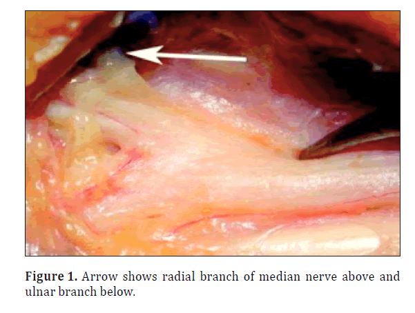
The carpal tunnel was exposed utilizing a standard S-shaped incision. It was clearly noted that median nerve was divided into it’s radial and ulnar branches approximately 2.5cm proximal to the flexor crease of the wrist. Both these branches entered the carpal tunnel but the radial branch was 0.5 cm deeper than the ulnar branch. Each branch was encased in a separate compartment comprised of cylindrical fibers originating from the transverse carpal ligament (Figure 1). The two compartments were 2 cm apart in the coronal plane. Each compartment was decompressed individually until the nerves were free. There was no communicating branch between the radial and ulnar divisions. The ulnar compartment was significantly less capacious when compared to the radial compartment.
Decompression of both these compartments resulted in complete resolution of the patient’s symptoms.
Discussion
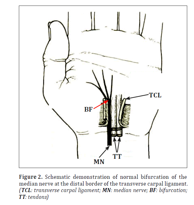
The terminal branches of the median nerve usually divide at the distal border of the transverse carpal ligament (Figure 2).
The median nerve rarely bifurcates in the flexor forearm. There have been only a few reports however of the median nerve dividing into two branches at the distal third of the forearm [5,6]. In the majority of the cases reported, both divisions of the median nerve entered the palm beneath the transverse carpal ligament.
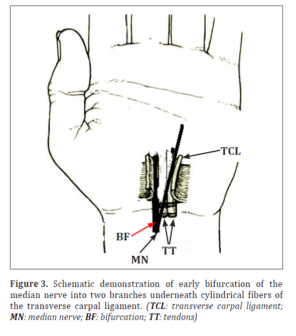
As far as the authors are aware, this is only the third documented case of a bifid median nerve with a double compartment [1,7]. We believe that this is the first case that demonstrated two cylindrical compartments arising from the transverse carpal ligament which encased each individual branch (Figure 3).
We opine that this is important to report as surgeons need to be wary of other variants of the median nerve when performing a carpal tunnel release. High bifurcation and compartmentalization of the median nerve described in our case may have an important bearing on the vulnerability of the nerve during surgical decompression.
It may also be important to note the branches of the nerve that may need separate decompression with the goal of symptom alleviation.
References
- Amadio PC. Bifid median nerve with a double compartment within the transverse carpal canal. J Hand Surg Am. 1987; 12: 366–368.
- Crandall RC, Hamel AL. Bipartite median nerve at the wrist. Report of a case. J Bone Joint Surg Am. 1979; 61: 311.
- Lanz U. Anatomical variations of the median nerve in the carpal tunnel. J Hand Surg Am. 1977; 2: 44–53.
- Falconer D, Spinner M. Anatomic variations in the motor and sensory supply of the thumb. Clin Orthop Relat Res. 1985; 195: 83–96.
- Eiken O, Carstam N, Eddeland A. Anomalous distal branching of the median nerve. Case reports. Scand J Plast Reconstr Surg. 1971; 5: 149–152.
- Kessler I. Unusual distribution of the median nerve at the wrist. A case report. Clin Orthop Relat Res. 1969; 67: 124–126.
- Takami H, Takahashi S, Ando M. Bipartite median nerve with a double compartment within the transverse carpal canal. Arch Orthop Trauma Surg. 2001; 121: 230–231.
Ameya S. Kamat* and Kelvin Woon
Department of Neurosurgery, Wellington Regional Hospital, Wellington, New Zealand
- *Corresponding Author:
- Dr. Ameya S. Kamat
Department of Neurosurgery, Wellington Regional Hospital, Riddiford St. Wellington South, Wellington, New Zealand
Tel: +64 4 385 5999
E-mail: amskam@gmail.com
Date of Received: October 3rd, 2012
Date of Accepted: August 28th, 2013
Published Online: June 1st, 2014
© Int J Anat Var (IJAV). 2014; 7: 24–26.
Abstract
The median nerve courses through the carpal tunnel before entering the hand. Numerous descriptions of this pathway exist. Several variants exist and have been documented. We report a case of an unusually high bifurcation of the median nerve into radial and ulnar branches proximal to the transverse carpal ligament. Another factor that makes our case unique is that both the branches were encapsulated by fibers arising from the transverse carpal ligament and were separated into two separate compartments within this ligament.
-Keywords
median nerve, carpal tunnel, bifurcation
Introduction
The median nerve courses through the carpal tunnel before entering the hand. Numerous descriptions of this pathway exist [1,2], which state that the terminal branches of the median nerve bifurcate into radial and ulnar divisions, at the distal border of the transverse carpal ligament.
Several variants exist and have been documented. Some variations describe the course of the thenar branch, the accessory branches at the distal carpal tunnel, high division of the median nerve and accessory branches proximal to the carpal tunnel [3,4].
We report a case of an unusually high bifurcation of the median nerve into radial and ulnar branches proximal to the transverse carpal ligament. Another factor that makes our case unique is that both the branches were encapsulated by fibers arising from the transverse carpal ligament and were separated into two separate compartments within this ligament.
Case Report
We describe the case of a 56-year-old, right hand dominant man who presented with tingling in the thumb, index, middle and the lateral half of his ring finger in the right hand for 3 years duration. Examination demonstrated mild thenar wasting and numbness over his thenar eminence. Further testing revealed and a positive Tinel’s sign and Phalen’s test. The Straight Arm Raise test was also positive. There was no motor weakness on examination.
Nerve conduction studies revealed median nerve entrapment in the carpal tunnel. He was hence brought forward for a carpal tunnel release under local anesthetic using Marcaine 0.25% with adrenaline.
The carpal tunnel was exposed utilizing a standard S-shaped incision. It was clearly noted that median nerve was divided into it’s radial and ulnar branches approximately 2.5cm proximal to the flexor crease of the wrist. Both these branches entered the carpal tunnel but the radial branch was 0.5 cm deeper than the ulnar branch. Each branch was encased in a separate compartment comprised of cylindrical fibers originating from the transverse carpal ligament (Figure 1). The two compartments were 2 cm apart in the coronal plane. Each compartment was decompressed individually until the nerves were free. There was no communicating branch between the radial and ulnar divisions. The ulnar compartment was significantly less capacious when compared to the radial compartment.
Decompression of both these compartments resulted in complete resolution of the patient’s symptoms.
Discussion
The terminal branches of the median nerve usually divide at the distal border of the transverse carpal ligament (Figure 2).
The median nerve rarely bifurcates in the flexor forearm. There have been only a few reports however of the median nerve dividing into two branches at the distal third of the forearm [5,6]. In the majority of the cases reported, both divisions of the median nerve entered the palm beneath the transverse carpal ligament.
As far as the authors are aware, this is only the third documented case of a bifid median nerve with a double compartment [1,7]. We believe that this is the first case that demonstrated two cylindrical compartments arising from the transverse carpal ligament which encased each individual branch (Figure 3).
We opine that this is important to report as surgeons need to be wary of other variants of the median nerve when performing a carpal tunnel release. High bifurcation and compartmentalization of the median nerve described in our case may have an important bearing on the vulnerability of the nerve during surgical decompression.
It may also be important to note the branches of the nerve that may need separate decompression with the goal of symptom alleviation.
References
- Amadio PC. Bifid median nerve with a double compartment within the transverse carpal canal. J Hand Surg Am. 1987; 12: 366–368.
- Crandall RC, Hamel AL. Bipartite median nerve at the wrist. Report of a case. J Bone Joint Surg Am. 1979; 61: 311.
- Lanz U. Anatomical variations of the median nerve in the carpal tunnel. J Hand Surg Am. 1977; 2: 44–53.
- Falconer D, Spinner M. Anatomic variations in the motor and sensory supply of the thumb. Clin Orthop Relat Res. 1985; 195: 83–96.
- Eiken O, Carstam N, Eddeland A. Anomalous distal branching of the median nerve. Case reports. Scand J Plast Reconstr Surg. 1971; 5: 149–152.
- Kessler I. Unusual distribution of the median nerve at the wrist. A case report. Clin Orthop Relat Res. 1969; 67: 124–126.
- Takami H, Takahashi S, Ando M. Bipartite median nerve with a double compartment within the transverse carpal canal. Arch Orthop Trauma Surg. 2001; 121: 230–231.


