Variant origin and course of ulnar artery – a case report
Surekha D Shetty, Satheesha Nayak B*, Ashwini Ls, Srinivasa Rao Sirasanagandla
Department of Anatomy, Melaka Manipal Medical College (Manipal Campus), Manipal University, Madhav Nagar, Manipal, Karnataka State, India.
- *Corresponding Author:
- Dr. Satheesha Nayak B
Professor and Head, Department of Anatomy, MMMC (Manipal Campus), International Centre for Health Sci., Manipal University, Madhav Nagar, Manipal, Udupi District, Karnataka State, 576 104, India.
Tel: +91 820 2922519
E-mail: nayaksathish@yahoo.com
Date of Received: February 23rd, 2012
Date of Accepted: October 18th, 2012
Published Online: February 14th, 2013
© Int J Anat Var (IJAV). 2013; 6: 45–46.
[ft_below_content] =>Keywords
ulnar artery, brachial artery, common interosseous artery, anatomical variations, upper limb
Introduction
Variations of the arterial patterns in the upper limb have been the subject of many anatomical studies due to their high incidence. Brachial artery is the main artery of the arm. It is a continuation of axillary artery at the lower border of teres major muscle. It usually terminates at the level of neck of radius in the cubital fossa by dividing into radial and ulnar arteries. The radial artery runs along the lateral part of the front of the forearm with the superficial branch of radial nerve. The ulnar artery passes medially deep to the pronator teres muscle and then runs to the distal part of the forearm together with the ulnar nerve. Superficial ulnar artery is a rare variation of the ulnar artery. It usually arises higher up, either in the axilla or the arm and runs a superficial course in the forearm before entering the hand [1].
Case Report
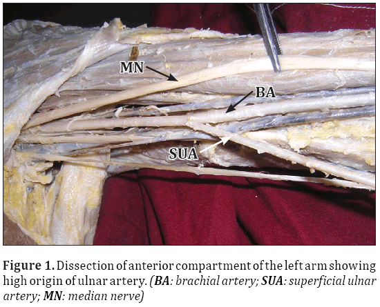
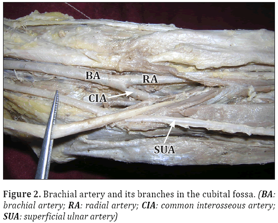
Variant origin and course of ulnar artery were observed during routine dissection of an adult male cadaver aged approximately 50 years. This variation was noted in the left upper limb. The ulnar artery originated from brachial artery in the proximal part of the arm and ran downwards parallel to median nerve, just deep to the brachial fascia to reach the cubital fossa (Figure 1). Then, it passed deep to the palmaris longus tendon in the forearm. At the wrist, it coursed in front of the flexor retinaculum to enter the palm. The brachial artery had a usual course till the cubital fossa. At the cubital fossa the brachial artery divided into two branches; a common interossoeus artery and a radial artery (Figure 2). The radial and common interosseous arteries had a usual course and branching pattern. But the common interosseous artery was much larger than usual.
Discussion
Knowledge of origin, course and distribution of ulnar artery is important due to its clinical implications. Anatomic variations in the major arteries of the upper extremities have been reported in 11-24.4% of individuals [2]. Presence of unusual blood vessels may be due to the persistence of vessels that normally get obliterated during the process of development [3]. The overall incidence of the superficial ulnar artery varies between 0.67% and 9.38% as reported in the various studies conducted throughout the world. The reported incidence of the superficial ulnar artery arising from the axillary artery varies between 0.7% and 2% [1]. The bilateral presence of the superficial ulnar artery with a different origin on each side is even more rare [4]. When the superficial ulnar artery is present, the brachial artery commonly terminates as the radial and common interosseous arteries [5]. High origin of superficial ulnar artery has been reported by Bozer et al., [6]. The superficial ulnar artery may be associated with the absence of the palmaris longus muscle. The development and clinical significance of superficial ulnar artery has also been reported by Reddy and Vollala [7].
Pulakunta et al., have reported a variation of co-existence of superficial ulnar artery and aneurysm of the deep palmar arch in the hand [8]. Sieg et al., and Fadel et al., have reported superficial ulnar artery and its clinical importance [9,10]. The superficial ulnar artery arises frequently from the lower third of the brachial artery, less frequently from the upper third and rarely from the middle third. In the present case the superficial ulnar artery arose from the junction of the upper and middle third of the brachial artery. Sanudo et al., [11] reported a case where superficial ulnar artery anastomosed with a larger anterior interosseous artery to supply the wrist and hand. Krishnamurthy et al., have also reported a high origin and superficial course of ulnar artery [12]. Presence of bilateral superficial ulnar arteries with an unusual arch in the forearm has been reported by Shankar et al. [13].
A superficial ulnar artery may complicate intravenous drug administration, venipuncture, and percutaneous brachial catheterization. Superficial course of ulnar artery from the middle of the arm till distal part of the forearm makes it vulnerable for injuries. Any superficial cut might result in severe bleeding. Knowledge of this variation is very important to radiologists, orthopedic & plastic surgeons for appropriately planning the operative procedures.
References
- Natsis K, Papadopoulou AL, Paraskevas G, Totlis T, Tsikaras P. High origin of a superficial ulnar artery arising from the axillary artery : anatomy, embryology, clinical significance and a review of the literature. Folia Morphol (Warsz). 2006; 65: 400–405.
- Uglietta JB, Kadir S. Arteriographic study of variant arterial anatomy of variant arterial anatomy of the upper extremities. Cardiovasc Intervent Radiol. 1989; 12: 145–148.
- Arey LB. Developmental Anatomy. 6th Ed., W.B. Philadelphia, Saunders Co. 1957; 375–377.
- Yazar F, Kirici Y, Ozan H, Aldur MM. An unusual variation of the superficial ulnar artery. Surg Radiol Anat. 1999; 21: 155–157.
- Rodriguez-Niedenfuhr M, Vazquez T, Nearn L, Ferreira B, Parkin I, Sanudo JR. Variations of the arterial pattern in the upper limb revisited: a morphological and statistical study, with a review of the literature. J Anat. 2001; 199: 547–566.
- Bozer C, Ulucam E, Kutoglu T. A case of originated high superficial ulnar artery. Trakia Journal of sciences. 2004; 2: 70–73.
- Reddy S, Vollala VR. The superficial ulnar artery: development and clinical significance. J Vasc Bras. 2007; 6: 285–288.
- Pulakunta T, Potu BK, Vollala VR, Goranta VR, Thomas H. Co-existence of superficial ulnar artery and aneurysm of the deep palmar arch in the hand. Bratisl Lek Listy. 2009; 110: 738–739.
- Sieg P, Jacobsen HC, Hakim SG, Hermes D. Superficial ulnar artery: curse or blessing in harvesting fasciocutaneous forearm flaps. Head Neck. 2006; 28: 447–452.
- Fadel RA, Amonoo-Kuofi HS. The superficial ulnar artery: development and surgical significance. Clin Anat. 1996; 9: 128–132.
- Sañudo JR, Mirapeix RM, Garcia R, Rodriguez-Nidenfünr M. Superficial ulnar artery anastomosing with a larger anterior interosseous artery to supply the wrist and hand. J Anat. 1998; 192: 439–441.
- Krishnamurthy A, Kumar M, Nayak SR, Prabhu LV. High origin and superficial course of ulnar artery: a case report. Firat Tip Dergisi. 2006; 11: 66–67.
- Shankar N, Veeramani R. Bilateral superficial ulnar arteries with an unusual arch in the forearm. Int J Anat Var (IJAV). 2009; 2: 24–26.
Surekha D Shetty, Satheesha Nayak B*, Ashwini Ls, Srinivasa Rao Sirasanagandla
Department of Anatomy, Melaka Manipal Medical College (Manipal Campus), Manipal University, Madhav Nagar, Manipal, Karnataka State, India.
- *Corresponding Author:
- Dr. Satheesha Nayak B
Professor and Head, Department of Anatomy, MMMC (Manipal Campus), International Centre for Health Sci., Manipal University, Madhav Nagar, Manipal, Udupi District, Karnataka State, 576 104, India.
Tel: +91 820 2922519
E-mail: nayaksathish@yahoo.com
Date of Received: February 23rd, 2012
Date of Accepted: October 18th, 2012
Published Online: February 14th, 2013
© Int J Anat Var (IJAV). 2013; 6: 45–46.
Abstract
Variations in the arterial system of the upper limb are well documented. A thorough knowledge of arteries of the upper extremity is necessary during vascular and reconstructive surgery and also in evaluation of angiographic images. We present here a case of variant origin and course of ulnar artery. The ulnar artery had a high origin from the brachial artery in the middle of the arm and proceeded superficially in the forearm but had a normal termination in the hand. The brachial artery had a usual course in the arm but in the cubital fossa it divided into the radial and common interosseous arteries. Knowledge of this variation is important to the radiologists, orthopedic and plastic surgeons for appropriate planning of operative procedures involving superficial ulnar artery.
-Keywords
ulnar artery, brachial artery, common interosseous artery, anatomical variations, upper limb
Introduction
Variations of the arterial patterns in the upper limb have been the subject of many anatomical studies due to their high incidence. Brachial artery is the main artery of the arm. It is a continuation of axillary artery at the lower border of teres major muscle. It usually terminates at the level of neck of radius in the cubital fossa by dividing into radial and ulnar arteries. The radial artery runs along the lateral part of the front of the forearm with the superficial branch of radial nerve. The ulnar artery passes medially deep to the pronator teres muscle and then runs to the distal part of the forearm together with the ulnar nerve. Superficial ulnar artery is a rare variation of the ulnar artery. It usually arises higher up, either in the axilla or the arm and runs a superficial course in the forearm before entering the hand [1].
Case Report
Variant origin and course of ulnar artery were observed during routine dissection of an adult male cadaver aged approximately 50 years. This variation was noted in the left upper limb. The ulnar artery originated from brachial artery in the proximal part of the arm and ran downwards parallel to median nerve, just deep to the brachial fascia to reach the cubital fossa (Figure 1). Then, it passed deep to the palmaris longus tendon in the forearm. At the wrist, it coursed in front of the flexor retinaculum to enter the palm. The brachial artery had a usual course till the cubital fossa. At the cubital fossa the brachial artery divided into two branches; a common interossoeus artery and a radial artery (Figure 2). The radial and common interosseous arteries had a usual course and branching pattern. But the common interosseous artery was much larger than usual.
Discussion
Knowledge of origin, course and distribution of ulnar artery is important due to its clinical implications. Anatomic variations in the major arteries of the upper extremities have been reported in 11-24.4% of individuals [2]. Presence of unusual blood vessels may be due to the persistence of vessels that normally get obliterated during the process of development [3]. The overall incidence of the superficial ulnar artery varies between 0.67% and 9.38% as reported in the various studies conducted throughout the world. The reported incidence of the superficial ulnar artery arising from the axillary artery varies between 0.7% and 2% [1]. The bilateral presence of the superficial ulnar artery with a different origin on each side is even more rare [4]. When the superficial ulnar artery is present, the brachial artery commonly terminates as the radial and common interosseous arteries [5]. High origin of superficial ulnar artery has been reported by Bozer et al., [6]. The superficial ulnar artery may be associated with the absence of the palmaris longus muscle. The development and clinical significance of superficial ulnar artery has also been reported by Reddy and Vollala [7].
Pulakunta et al., have reported a variation of co-existence of superficial ulnar artery and aneurysm of the deep palmar arch in the hand [8]. Sieg et al., and Fadel et al., have reported superficial ulnar artery and its clinical importance [9,10]. The superficial ulnar artery arises frequently from the lower third of the brachial artery, less frequently from the upper third and rarely from the middle third. In the present case the superficial ulnar artery arose from the junction of the upper and middle third of the brachial artery. Sanudo et al., [11] reported a case where superficial ulnar artery anastomosed with a larger anterior interosseous artery to supply the wrist and hand. Krishnamurthy et al., have also reported a high origin and superficial course of ulnar artery [12]. Presence of bilateral superficial ulnar arteries with an unusual arch in the forearm has been reported by Shankar et al. [13].
A superficial ulnar artery may complicate intravenous drug administration, venipuncture, and percutaneous brachial catheterization. Superficial course of ulnar artery from the middle of the arm till distal part of the forearm makes it vulnerable for injuries. Any superficial cut might result in severe bleeding. Knowledge of this variation is very important to radiologists, orthopedic & plastic surgeons for appropriately planning the operative procedures.
References
- Natsis K, Papadopoulou AL, Paraskevas G, Totlis T, Tsikaras P. High origin of a superficial ulnar artery arising from the axillary artery : anatomy, embryology, clinical significance and a review of the literature. Folia Morphol (Warsz). 2006; 65: 400–405.
- Uglietta JB, Kadir S. Arteriographic study of variant arterial anatomy of variant arterial anatomy of the upper extremities. Cardiovasc Intervent Radiol. 1989; 12: 145–148.
- Arey LB. Developmental Anatomy. 6th Ed., W.B. Philadelphia, Saunders Co. 1957; 375–377.
- Yazar F, Kirici Y, Ozan H, Aldur MM. An unusual variation of the superficial ulnar artery. Surg Radiol Anat. 1999; 21: 155–157.
- Rodriguez-Niedenfuhr M, Vazquez T, Nearn L, Ferreira B, Parkin I, Sanudo JR. Variations of the arterial pattern in the upper limb revisited: a morphological and statistical study, with a review of the literature. J Anat. 2001; 199: 547–566.
- Bozer C, Ulucam E, Kutoglu T. A case of originated high superficial ulnar artery. Trakia Journal of sciences. 2004; 2: 70–73.
- Reddy S, Vollala VR. The superficial ulnar artery: development and clinical significance. J Vasc Bras. 2007; 6: 285–288.
- Pulakunta T, Potu BK, Vollala VR, Goranta VR, Thomas H. Co-existence of superficial ulnar artery and aneurysm of the deep palmar arch in the hand. Bratisl Lek Listy. 2009; 110: 738–739.
- Sieg P, Jacobsen HC, Hakim SG, Hermes D. Superficial ulnar artery: curse or blessing in harvesting fasciocutaneous forearm flaps. Head Neck. 2006; 28: 447–452.
- Fadel RA, Amonoo-Kuofi HS. The superficial ulnar artery: development and surgical significance. Clin Anat. 1996; 9: 128–132.
- Sañudo JR, Mirapeix RM, Garcia R, Rodriguez-Nidenfünr M. Superficial ulnar artery anastomosing with a larger anterior interosseous artery to supply the wrist and hand. J Anat. 1998; 192: 439–441.
- Krishnamurthy A, Kumar M, Nayak SR, Prabhu LV. High origin and superficial course of ulnar artery: a case report. Firat Tip Dergisi. 2006; 11: 66–67.
- Shankar N, Veeramani R. Bilateral superficial ulnar arteries with an unusual arch in the forearm. Int J Anat Var (IJAV). 2009; 2: 24–26.


