Variant position of the facial nerve in parotid gland
Rajesh B. Astik*, Urvi H. Dave and Krishna Swami Gajendra
Department of Anatomy, GSL Medical College, Rajahmundry, District- East Godavari, Andhra Pradesh, India
- *Corresponding Author:
- Dr. Rajesh B. Astik
Associate Professor, Department of Anatomy, GSL Medical College, NH-5, Rajahmundry, District- East Godavari, Andhra Pradesh, 533296, India
Tel: +91 883 2484999
E-mail: astikrajesh@yahoo.co.in
Date of Received: July 15th, 2010
Date of Accepted: January 4th, 2011
Published Online: January 14th, 2011
© IJAV. 2011; 4: 3–4.
[ft_below_content] =>Keywords
facial nerve, parotid gland, retromandibular vein, total parotidectomy
Introduction
The retromandibular vein is formed by union of the maxillary and superficial temporal veins in the parotid gland [1]. The facial nerve enters the posteromedial surface of the parotid gland and crosses superficial to external carotid artery and retromandibular vein and divided into the cervicofacial and temporofacial divisions in the parotid gland [2].
During surgery for removal of tumors from the parotid gland, facial nerve can be injured because of its variant position between the maxillary and superficial temporal veins as found in this case. Purpose of this paper is to reduce unexpected bleeding from superficial temporal vein during surgery and postoperative morbidity related to facial nerve paralysis.
Case Report
We described a variant position of the facial nerve in parotid gland of the left side during routine educational dissection of a 55-year-old male cadaver in the Department of Anatomy, GSL Medical College.
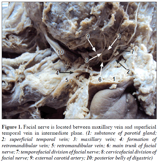
The main trunk of the facial nerve was located between the formative tributaries of the retromandibular vein, i.e., the maxillary and superficial temporal veins, and divided in temporofacial and cervicofacial divisions in between these two veins. These two divisions then crossed maxillary vein superficially instead of the retromandibular vein. The retromandibular vein was formed by union of maxillary and superficial temporal veins below the apex of parotid gland (Figure 1).
Figure 1: Facial nerve is located between maxillary vein and superficial temporal vein in intermediate plane. (1: substance of parotid gland; 2: superficial temporal vein; 3: maxillary vein; 4: formation of retromandibular vein; 5: retromandibular vein; 6: main trunk of facial nerve; 7: temporofacial division of facial nerve; 8: cervicofacial division of facial nerve; 9: external carotid artery; 10: posterior belly of digastric)
The branching pattern and distribution of the facial nerve and division of retromandibular vein were found as per described in the standard textbooks of anatomy.
Discussion
The risk of damage to the facial nerve during surgical procedures of the parotid gland revealed the importance of knowledge of detailed anatomy of this region [3].
In 90% of the cases the retromandibular vein was located on the medial side of the temporofacial and cervicofacial divisions of the facial nerve and in 10% the course of the retromandibular vein was lateral to the cervicofacial and medial to the temporofacial divisions [4]. Dingman et al. stated that in 98% cases, the retromandibular vein coursed medial to the mandibular branch of the facial nerve and in only 2% it coursed lateral to it [5]. Savary et al. reported that the cervicofacial division passed the superficial side of the retromandibular vein in all cases [6]. We found main trunk of the facial nerve and its divisions were forked between the maxillary and superficial temporal veins, so this variation could be used as a reference in surgery.
Retromandibular vein was a sensitive marker for identifying the location of parotid gland neoplasm with respect to the facial nerve on cross-sectional imaging [7]. The identification of the facial nerve in the parotid gland and its relation with either retromandibular vein or superficial temporal vein during parotidectomy or repair of facial trauma is a paradigmatic procedure [8]. The superficial temporal and retromandibular veins have been reported to be used as guide to expose facial nerve branches in the parotid gland in cases of open reduction of mandibular condyle fractures and also for superficial parotidectomy [9]. These veins were usually grafted into the carotid during endarterectomy and for surgery involving microvascular anastomosis especially in oral reconstruction procedures [10]. So knowledge of such variation is very important for surgeon during surgery to prevent unexpected bleeding from superficial temporal vein while dealing with the facial nerve.
Conclusion
Knowledge of this type of variant position of the facial nerve is important for the physicians as the facial nerve might be compressed by increased venous return from superficial temporal and maxillary veins as the nerve was forked between these two veins, and for the surgeons in order to avoid any intraoperative trial and error procedures which might lead to unexpected bleeding from the superficial temporal vein and facial nerve damage.
Acknowledgements
Our sincere thanks to all the people who helped and supported during the writing of this manuscript. We would thank our institution for allowing us to dissect cadaver and faculty members without whom this manuscript would have been a distant reality.
References
- Williams PL, Bannister LH, Berry MM, Collins P, Dyson M, Dussek JE, Ferguson MWJ. Gray’s Anatomy. 38th Ed., Edinburgh London, Churchill Livingstone. 1995: 1691.
- Potgieter W, Meiring JH, Boon JM, Pretorius E, Pretorius JP, Becker PJ. Mandibular landmarks as an aid in minimizing injury to the marginal mandibular branch: A metric and geometric anatomical study. Clin Anat. 2005; 18: 171–178.
- Laing MR, McKerrow WS. Intraparotid anatomy of the facial nerve and retromandibular vein. Br J Surg. 1988; 75: 310–312.
- Kopuz C, Ilgi S, Yavuz S, Onderoglu S. The morphology of the retromandibular vein in relation to the facial nerve in the parotid gland. Turk J Med Res. 1993; 11: 62–65.
- Dingman RO, Grabb WC. Surgical anatomy of the mandibular ramus of the facial nerve based on the dissection of 100 facial halves. Plast Reconstr Surg Transplant Bull. 1962; 29: 266–272.
- Savary V, Robert R, Rogez JM, Armstrong O, Leborgne J. The mandibular marginal ramus of the facial nerve: An anatomic and clinical study. Surg Radiol Anat. 1997; 19: 69–72.
- Divi V, Fatt MA, Teknos TN, Mukherji SK. Use of cross-sectional imaging in predicting surgical location of parotid neoplasms. J Comput Assist Tomogr. 2005; 29: 315–319.
- Pereira JA, Meri A, Potau JM, Prats-Galino A, Sancho JJ, Sitges-Serra A. A simple method for safe identification of the facial nerve using palpable landmarks. Arch Surg. 2004; 139: 745–747.
- Kawakami S, Tsukada S, Taniguchi W. The superficial temporal and retromandibular veins as guides to expose the facial nerve branches. Ann Plast Surg. 1994; 32: 295–299.
- Sabharwal P, Mukherjee D. Autogenous common facial vein or external jugular vein patch for carotid endarterectomy. Cardiovasc Surg. 1998; 6: 594–597.
Rajesh B. Astik*, Urvi H. Dave and Krishna Swami Gajendra
Department of Anatomy, GSL Medical College, Rajahmundry, District- East Godavari, Andhra Pradesh, India
- *Corresponding Author:
- Dr. Rajesh B. Astik
Associate Professor, Department of Anatomy, GSL Medical College, NH-5, Rajahmundry, District- East Godavari, Andhra Pradesh, 533296, India
Tel: +91 883 2484999
E-mail: astikrajesh@yahoo.co.in
Date of Received: July 15th, 2010
Date of Accepted: January 4th, 2011
Published Online: January 14th, 2011
© IJAV. 2011; 4: 3–4.
Abstract
The division of the parotid gland into superficial and deep lobes by facial nerve has an important implication in parotid gland neoplasm. This plane is used in superficial or total parotidectomy to avoid damage to the facial nerve. During routine dissection in the Department of Anatomy, we found variably located facial nerve in the parotid gland of the left side. The main trunk of the facial nerve was located between maxillary vein and superficial temporal vein. It was divided into temporofacial and cervicofacial divisions. Both divisions crossed maxillary vein superficially instead of retromandibular vein which was formed outside the parotid gland substance. The operating surgeon should be familiar with this variation during parotidectomy to reduce the iatrogenic injury to the facial nerve.
-Keywords
facial nerve, parotid gland, retromandibular vein, total parotidectomy
Introduction
The retromandibular vein is formed by union of the maxillary and superficial temporal veins in the parotid gland [1]. The facial nerve enters the posteromedial surface of the parotid gland and crosses superficial to external carotid artery and retromandibular vein and divided into the cervicofacial and temporofacial divisions in the parotid gland [2].
During surgery for removal of tumors from the parotid gland, facial nerve can be injured because of its variant position between the maxillary and superficial temporal veins as found in this case. Purpose of this paper is to reduce unexpected bleeding from superficial temporal vein during surgery and postoperative morbidity related to facial nerve paralysis.
Case Report
We described a variant position of the facial nerve in parotid gland of the left side during routine educational dissection of a 55-year-old male cadaver in the Department of Anatomy, GSL Medical College.
The main trunk of the facial nerve was located between the formative tributaries of the retromandibular vein, i.e., the maxillary and superficial temporal veins, and divided in temporofacial and cervicofacial divisions in between these two veins. These two divisions then crossed maxillary vein superficially instead of the retromandibular vein. The retromandibular vein was formed by union of maxillary and superficial temporal veins below the apex of parotid gland (Figure 1).
Figure 1: Facial nerve is located between maxillary vein and superficial temporal vein in intermediate plane. (1: substance of parotid gland; 2: superficial temporal vein; 3: maxillary vein; 4: formation of retromandibular vein; 5: retromandibular vein; 6: main trunk of facial nerve; 7: temporofacial division of facial nerve; 8: cervicofacial division of facial nerve; 9: external carotid artery; 10: posterior belly of digastric)
The branching pattern and distribution of the facial nerve and division of retromandibular vein were found as per described in the standard textbooks of anatomy.
Discussion
The risk of damage to the facial nerve during surgical procedures of the parotid gland revealed the importance of knowledge of detailed anatomy of this region [3].
In 90% of the cases the retromandibular vein was located on the medial side of the temporofacial and cervicofacial divisions of the facial nerve and in 10% the course of the retromandibular vein was lateral to the cervicofacial and medial to the temporofacial divisions [4]. Dingman et al. stated that in 98% cases, the retromandibular vein coursed medial to the mandibular branch of the facial nerve and in only 2% it coursed lateral to it [5]. Savary et al. reported that the cervicofacial division passed the superficial side of the retromandibular vein in all cases [6]. We found main trunk of the facial nerve and its divisions were forked between the maxillary and superficial temporal veins, so this variation could be used as a reference in surgery.
Retromandibular vein was a sensitive marker for identifying the location of parotid gland neoplasm with respect to the facial nerve on cross-sectional imaging [7]. The identification of the facial nerve in the parotid gland and its relation with either retromandibular vein or superficial temporal vein during parotidectomy or repair of facial trauma is a paradigmatic procedure [8]. The superficial temporal and retromandibular veins have been reported to be used as guide to expose facial nerve branches in the parotid gland in cases of open reduction of mandibular condyle fractures and also for superficial parotidectomy [9]. These veins were usually grafted into the carotid during endarterectomy and for surgery involving microvascular anastomosis especially in oral reconstruction procedures [10]. So knowledge of such variation is very important for surgeon during surgery to prevent unexpected bleeding from superficial temporal vein while dealing with the facial nerve.
Conclusion
Knowledge of this type of variant position of the facial nerve is important for the physicians as the facial nerve might be compressed by increased venous return from superficial temporal and maxillary veins as the nerve was forked between these two veins, and for the surgeons in order to avoid any intraoperative trial and error procedures which might lead to unexpected bleeding from the superficial temporal vein and facial nerve damage.
Acknowledgements
Our sincere thanks to all the people who helped and supported during the writing of this manuscript. We would thank our institution for allowing us to dissect cadaver and faculty members without whom this manuscript would have been a distant reality.
References
- Williams PL, Bannister LH, Berry MM, Collins P, Dyson M, Dussek JE, Ferguson MWJ. Gray’s Anatomy. 38th Ed., Edinburgh London, Churchill Livingstone. 1995: 1691.
- Potgieter W, Meiring JH, Boon JM, Pretorius E, Pretorius JP, Becker PJ. Mandibular landmarks as an aid in minimizing injury to the marginal mandibular branch: A metric and geometric anatomical study. Clin Anat. 2005; 18: 171–178.
- Laing MR, McKerrow WS. Intraparotid anatomy of the facial nerve and retromandibular vein. Br J Surg. 1988; 75: 310–312.
- Kopuz C, Ilgi S, Yavuz S, Onderoglu S. The morphology of the retromandibular vein in relation to the facial nerve in the parotid gland. Turk J Med Res. 1993; 11: 62–65.
- Dingman RO, Grabb WC. Surgical anatomy of the mandibular ramus of the facial nerve based on the dissection of 100 facial halves. Plast Reconstr Surg Transplant Bull. 1962; 29: 266–272.
- Savary V, Robert R, Rogez JM, Armstrong O, Leborgne J. The mandibular marginal ramus of the facial nerve: An anatomic and clinical study. Surg Radiol Anat. 1997; 19: 69–72.
- Divi V, Fatt MA, Teknos TN, Mukherji SK. Use of cross-sectional imaging in predicting surgical location of parotid neoplasms. J Comput Assist Tomogr. 2005; 29: 315–319.
- Pereira JA, Meri A, Potau JM, Prats-Galino A, Sancho JJ, Sitges-Serra A. A simple method for safe identification of the facial nerve using palpable landmarks. Arch Surg. 2004; 139: 745–747.
- Kawakami S, Tsukada S, Taniguchi W. The superficial temporal and retromandibular veins as guides to expose the facial nerve branches. Ann Plast Surg. 1994; 32: 295–299.
- Sabharwal P, Mukherjee D. Autogenous common facial vein or external jugular vein patch for carotid endarterectomy. Cardiovasc Surg. 1998; 6: 594–597.


