Variation in the termination of musculocutaneous nerve
Huban R Thomas1*, Bhagath K Potu1, Kumar MR Bhat1, Binu Margaret2, Venu Madhav3 and Stelin Wersely4
1Department of Anatomy, Centre for Basic Sciences, Kasturba Medical College, India
2Department of Pediatric Nursing, Manipal College of Nursing, India
3Department of Anatomy, Melaka Manipal Medical College (Manipal Campus), India
4Manipal University, Manipal, Karnataka; Department of Anatomy, Sree Mookambika Institute of Medical Sciences, Kulasekharam, Tamil Nadu, India
- *Corresponding Author:
- Huban R Thomas, BPT, MSc
Department of Anatomy, Centre for Basic Sciences Kasturba Medical College, Manipal University Manipal, Karnataka, India
Tel: +91 820 2922327
E-mail: huban_anatomy@yahoo.co.in
Date of Received: October 16th, 2009
Date of Accepted: April 9th, 2010
Published Online: May 17th, 2010
© Int J Anat Var (IJAV). 2010; 3: 78–79.
[ft_below_content] =>Keywords
variation, abnormal branch, brachial plexus
Introduction
Brachial plexus is formed by the anterior primary rami of spinal nerves C5, C6, C7, C8 and T1. The fibers of the plexus may be joined by branches from the fourth cervical and second thoracic nerves. C5 and C6 roots join to form upper trunk. C7 root forms the middle trunk. C8 and T1 roots join to form lower trunk. Each trunk divides into ventral and dorsal divisions. Ventral division of the lower trunk forms medial cord. Dorsal divisions of all the three trunks join to forms posterior cord. Ventral divisions of upper and middle trunk joint to forms lateral cord. Musculocutaneous nerve (MCN) is the branch from the lateral cord of the brachial plexus. The nerve initially accompanies the axillary artery, pierces the coracobrachialis muscle, and then passes downwards between the biceps brachii and brachialis. It supplies coracobrachialis, biceps brachii and medial part of brachialis muscles. Below the elbow joint the nerve is continuous as the lateral cutaneous nerve of the forearm.
Case Report
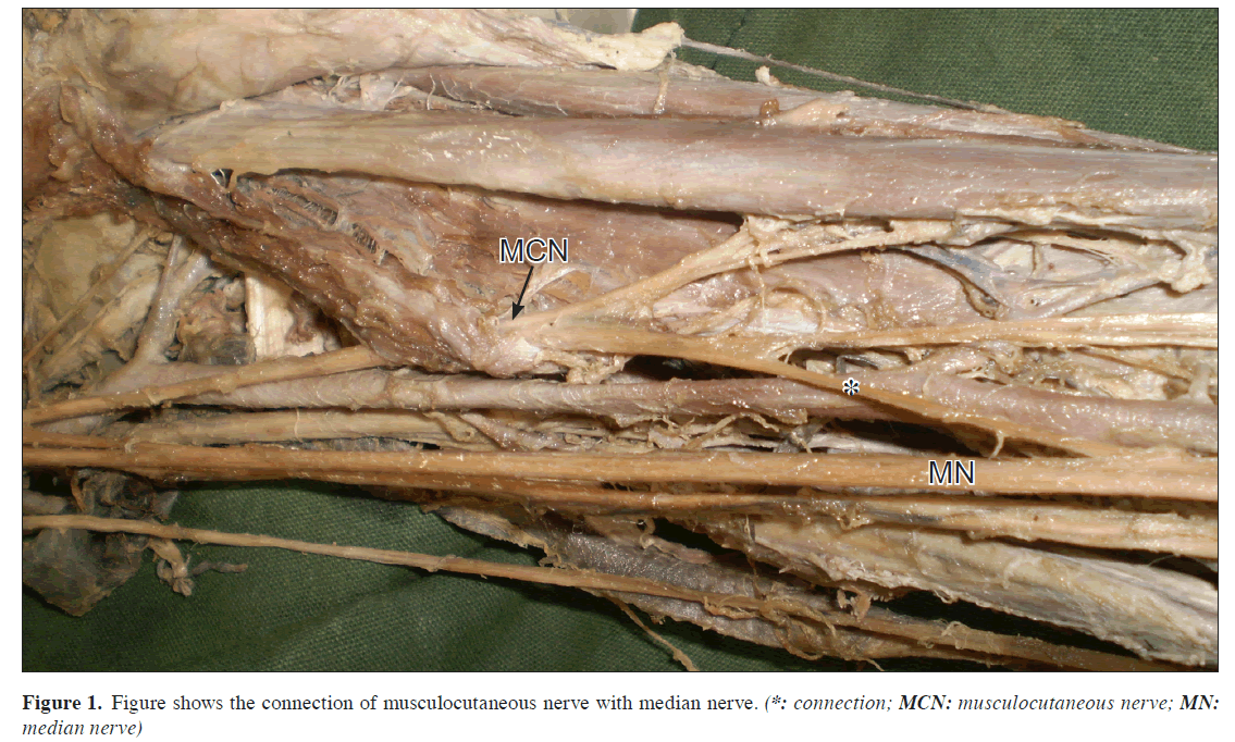
During routine educational dissection for medical undergraduates, we observed a variation in the termination of MCN in right upper limb of middle aged Indian male cadaver. After piercing coracobrachialis muscle, MCN divided into lateral cutaneous nerve of the forearm and another branch that joined the median nerve (MN) below the insertion of the coracobrachialis (Figure 1). Courses of all other major nerves like median, ulnar, axillary and radial were as usual.
Discussion
The present report localizes the anomalous termination of MCN in right upper limb of middle aged Indian male cadaver. There are different types of variations were reported during dissection of the brachial plexus. Venieratos and Anagnostopoulou reported three types of unusual communication between MN and MCN considering the coracobrachialis muscle as the reference point [1]. In type one, the communication was proximal to the entrance of the MCN into coracobrachialis. In type two, the communication was distal to the muscle. In type three, the nerve and the communicating branch did not pierce the muscle. Eglseder and Goldman noticed interconnections between the MCN and MN in 36% of dissections out of 54 cadavers [2]. Loukas and Aqueelah identify 4 different patterns of communication. Type I (54 communications, 45%): the communications were proximal to the point of entry of the MCN into the coracobrachialis; type II (42 communications, 35%): the communications were distal to the point of entry of the MCN into the coracobrachialis; type III (11 communications, 9%): the MCN did not pierce the coracobrachialis; and Type IV (9 communications, 8%) out of 129 formalin-fixed cadavers [3]. Venieratos and Anagnostopoulou found types of communications between the MCN and MN based on the sites of communication. Type I: the communication was proximal to the entrance of the musculocutaneous nerve into coracobrachialis; type II: the communication was distal to the muscle; type III: the nerve as well as the communicating branch did not pierce the muscle out of 79 cadavers [1]. Prasada Rao and Chaudhary reported eight instances of communication from MCN to the MN and bilateral communication in two cadavers [4]. Chauhan and Roy also reported unusual communication between the median and MCN in their case report [5]. We believe that this variation is because of some of the fibers of MN might have passed through the MCN and then join with MN after the nerve leaves the coracobrachialis muscle.
References
- Venieratos D, Anagnostopoulou S. Classification of communications between the musculocutaneous and median nerves. Clin Anat 1998; 11: 327–331.
- Eglseder WA Jr, Goldman M. Anatomic variations of the musculocutaneous nerve in the arm. Am J Orthop (Belle Mead NJ). 1997; 26: 777–780.
- Loukas M, Aqueelah H. Musculocutaneous and median nerve connections within, proximal and distal to the coracobrachialis muscle. Folia Morphol (Warsz). 2005; 64: 101–108.
- Prasada Rao PV, Chaudhary SC. Communication of the musculocutaneous nerve with the median nerve. East Afr Med J. 2000; 77: 498–503.
- Chauhan R, Roy TS. Communication between the median and musculocutaneous nerve – a case report. J Anat Soc India. 2002; 51: 72–75.
Huban R Thomas1*, Bhagath K Potu1, Kumar MR Bhat1, Binu Margaret2, Venu Madhav3 and Stelin Wersely4
1Department of Anatomy, Centre for Basic Sciences, Kasturba Medical College, India
2Department of Pediatric Nursing, Manipal College of Nursing, India
3Department of Anatomy, Melaka Manipal Medical College (Manipal Campus), India
4Manipal University, Manipal, Karnataka; Department of Anatomy, Sree Mookambika Institute of Medical Sciences, Kulasekharam, Tamil Nadu, India
- *Corresponding Author:
- Huban R Thomas, BPT, MSc
Department of Anatomy, Centre for Basic Sciences Kasturba Medical College, Manipal University Manipal, Karnataka, India
Tel: +91 820 2922327
E-mail: huban_anatomy@yahoo.co.in
Date of Received: October 16th, 2009
Date of Accepted: April 9th, 2010
Published Online: May 17th, 2010
© Int J Anat Var (IJAV). 2010; 3: 78–79.
Abstract
The present report describes a case of variation of the musculocutaneous nerve observed in a middle aged Indian male cadaver during routine educational dissection. We examined a variation in the termination of musculocutaneous nerve in right upper limb. After piercing coracobrachialis muscle musculocutaneous nerve divided into lateral cutaneous nerve of the forearm and another branch that joined with median nerve below the insertion of the coracobrachialis. This abnormal branch coming from the musculocutaneous nerve had a very close oblique course over the brachial artery. Precise knowledge of variations of this report may help to plan a surgery in the region of axilla and arm, traumatology of the shoulder joint and plastic and reconstructive repair operations.
-Keywords
variation, abnormal branch, brachial plexus
Introduction
Brachial plexus is formed by the anterior primary rami of spinal nerves C5, C6, C7, C8 and T1. The fibers of the plexus may be joined by branches from the fourth cervical and second thoracic nerves. C5 and C6 roots join to form upper trunk. C7 root forms the middle trunk. C8 and T1 roots join to form lower trunk. Each trunk divides into ventral and dorsal divisions. Ventral division of the lower trunk forms medial cord. Dorsal divisions of all the three trunks join to forms posterior cord. Ventral divisions of upper and middle trunk joint to forms lateral cord. Musculocutaneous nerve (MCN) is the branch from the lateral cord of the brachial plexus. The nerve initially accompanies the axillary artery, pierces the coracobrachialis muscle, and then passes downwards between the biceps brachii and brachialis. It supplies coracobrachialis, biceps brachii and medial part of brachialis muscles. Below the elbow joint the nerve is continuous as the lateral cutaneous nerve of the forearm.
Case Report
During routine educational dissection for medical undergraduates, we observed a variation in the termination of MCN in right upper limb of middle aged Indian male cadaver. After piercing coracobrachialis muscle, MCN divided into lateral cutaneous nerve of the forearm and another branch that joined the median nerve (MN) below the insertion of the coracobrachialis (Figure 1). Courses of all other major nerves like median, ulnar, axillary and radial were as usual.
Discussion
The present report localizes the anomalous termination of MCN in right upper limb of middle aged Indian male cadaver. There are different types of variations were reported during dissection of the brachial plexus. Venieratos and Anagnostopoulou reported three types of unusual communication between MN and MCN considering the coracobrachialis muscle as the reference point [1]. In type one, the communication was proximal to the entrance of the MCN into coracobrachialis. In type two, the communication was distal to the muscle. In type three, the nerve and the communicating branch did not pierce the muscle. Eglseder and Goldman noticed interconnections between the MCN and MN in 36% of dissections out of 54 cadavers [2]. Loukas and Aqueelah identify 4 different patterns of communication. Type I (54 communications, 45%): the communications were proximal to the point of entry of the MCN into the coracobrachialis; type II (42 communications, 35%): the communications were distal to the point of entry of the MCN into the coracobrachialis; type III (11 communications, 9%): the MCN did not pierce the coracobrachialis; and Type IV (9 communications, 8%) out of 129 formalin-fixed cadavers [3]. Venieratos and Anagnostopoulou found types of communications between the MCN and MN based on the sites of communication. Type I: the communication was proximal to the entrance of the musculocutaneous nerve into coracobrachialis; type II: the communication was distal to the muscle; type III: the nerve as well as the communicating branch did not pierce the muscle out of 79 cadavers [1]. Prasada Rao and Chaudhary reported eight instances of communication from MCN to the MN and bilateral communication in two cadavers [4]. Chauhan and Roy also reported unusual communication between the median and MCN in their case report [5]. We believe that this variation is because of some of the fibers of MN might have passed through the MCN and then join with MN after the nerve leaves the coracobrachialis muscle.
References
- Venieratos D, Anagnostopoulou S. Classification of communications between the musculocutaneous and median nerves. Clin Anat 1998; 11: 327–331.
- Eglseder WA Jr, Goldman M. Anatomic variations of the musculocutaneous nerve in the arm. Am J Orthop (Belle Mead NJ). 1997; 26: 777–780.
- Loukas M, Aqueelah H. Musculocutaneous and median nerve connections within, proximal and distal to the coracobrachialis muscle. Folia Morphol (Warsz). 2005; 64: 101–108.
- Prasada Rao PV, Chaudhary SC. Communication of the musculocutaneous nerve with the median nerve. East Afr Med J. 2000; 77: 498–503.
- Chauhan R, Roy TS. Communication between the median and musculocutaneous nerve – a case report. J Anat Soc India. 2002; 51: 72–75.


