Vasa aberrantia connecting the brachial and radial arteries
Devi Sankar K*, Sharmila Bhanu P, Susan PJ, Gajendra K
Department of Anatomy, Narayana Medical College, Chinthareddy Palem, Nellore, Andhra Pradesh, India.
- *Corresponding Author:
- Devi Sankar K, MSc
Assistant Professor, Department of Anatomy, Narayana Medical College, Chinthareddy Palem, Nellore, Andhra Pradesh, 524 002, India.
Tel: +91 949 0948006
E-mail: lesanshar@gmail.com
Date of Received: March 19th, 2009
Date of Accepted: July 10th, 2009
Published Online: July 13th, 2009
© IJAV. 2009; 2: 75–77.
[ft_below_content] =>Keywords
vasa aberrentia, anastomotic artery, brachial artery, radial artery
Introduction
The vascular variations of upper limb are more common and has its own significance in relation to the clinical and surgical procedures carried out in respective parts of the upper limb. Knowledge of the variations of arterial pattern of the upper limb has been thoroughly studied by many authors [1,2].
The brachial artery normally is the continuation of axillary artery from the lower border of teres major, courses in the anterior compartment of arm. In the arm, median nerve first lies anterior to the 3rd part of the axillary artery and then along its course it crosses the brachial artery anteriorly from lateral to medial side. As brachial artery enters the cubital fossa, at the level of neck of radius it divides into radial and ulnar artery. The formation, origin and distribution of blood vessels may vary at any level of upper limb during its development and it can be demonstrated on the developmental basis.
Few of the developmental vascular anomalies of upper limb are presence of vasa aberrantia, higher bifurcation brachial artery, superficial brachioradial artery, median artery of the forearm, and variations in the formation of superficial or deep palmar arch [3,4].
Case Report
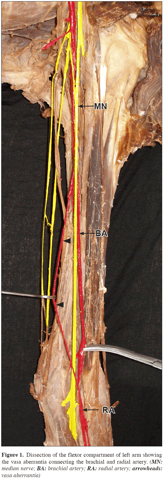
During routine dissections we observed an aberrant artery (vasa aberrantia) connecting the brachial artery to the radial artery in the left upper limb of a 52-year-old male cadaver. This vasa aberrantia was originating from the medial side of the brachial artery at the level of insertion of coracobrachialis, and was 12.8 cm long (Figure 1). At its origin, this aberrant artery was medial and posterior to the median nerve. In the lower third of the arm, this anastomotic branch crossed the median nerve anteriorly from medial to lateral side, and joined the radial artery on its lateral side. Along its course it gave branches to the muscles of the anterior compartment.
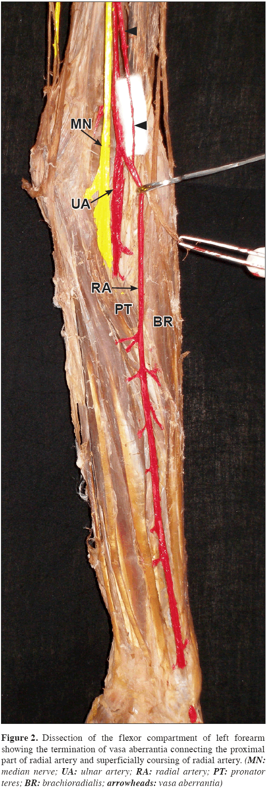
In the cubital fossa radial and ulnar arteries arose from the brachial artery, half a centimeter above the level of the medial epicondyle of humerus, instead of its usual origin at the level of neck of radius. The radial artery was superficial throughout its course, from its origin to the anatomical snuffbox and was represented as superficial radial artery (Figure 2).
Figure 2: Dissection of the flexor compartment of left forearm showing the termination of vasa aberrantia connecting the proximal part of radial artery and superficially coursing of radial artery. (MN: median nerve; UA: ulnar artery; RA: radial artery; PT: pronator teres; BR: brachioradialis; arrowheads: vasa aberrantia)
In the forearm, superficial radial artery ran superficial to the muscles of the forearm and deep to the antebrachial fascia along the radial border. When the artery reached the wrist, it was entrapped by the lateral fibers of flexor retinaculum, which converted itself into a compartment for the artery. The artery passed through this compartment and entered the hand deep to the thenar muscles to complete the superficial palmar arch. Throughout its course, this superficial radial artery supplied the flexor compartment muscles of forearm via numerous branches. The common interosseous and other anastomotic branches had usual origin and course. The brachial plexus and its branches did not show any variation.
Discussion
The vascular anatomy is a most inconstant one in the body, and variations are usually the result of unusual formation of blood vessels during development. These variations are mostly observed during the surgical/angiographic procedures or during cadaver dissections. The earliest studies on the variations of the arterial system were reported by Singer [5].
Developmentally the 7th cervical intersegmental artery gives rise to the axial artery that forms the major artery from which other branches of the upper limb arises. The proximal portion of axial artery above the level of teres major forms the axillary artery, and distal to it continues as brachial artery, and finally in the cubital fossa, as interosseous artery. The radial and ulnar arteries arise late in the development, because of the interosseous artery becomes reduced in size [5].
During development, the radial artery takes its origin from the brachial artery proximal to the ulnar artery. After the establishment of new communications with the main trunk, at or close to the level of origin of ulnar artery, the upper part of the radial artery regresses [6,7]. Failure of this original segment of radial artery to disappear, which appears first during development, persists as an anastomotic branch (vasa aberrantia) between the axillary or brachial and radial artery [4].
According to Rodriguez-Niedenfuhr et al., the development of arteries of the upper limb initially starts as a capillary plexus, which gradually enlarge, differentiate and regress to finally reach the adult type vascular pattern of the upper limb [8]. Variations in the formation of stages of this capillary plexus forming into definitive blood vessels gives rise to variations of the arterial pattern of the upper limb [2,3,5,9].
The vasa aberrantia, observed in the present case, represents the non-regressive proximal portion of the radial artery, which was formed at first during development. This anastomotic branch that rejoined radial artery on its lateral side may be taken as a confirmation that it is the remnant of the proximal part of radial artery. The vasa aberrentia may also persist between the axillary and radial arteries [4].
Numerous arterial variations have been reported by many authors, of which the most frequently observed one is the higher bifurcation of brachial artery from brachial or axillary artery. The incidence of higher origin of radial artery from axillary artery was reported as 2.13% [10]. The superficial radial artery from brachial artery was found to be 14.26% in cadaveric studies and 9.75% in angiographic studies [11].
The presence of aberrant arteries of the upper limb as we encountered in the present case, may alter the evaluation of angiographic images. It may be injured during venipuncture and is vulnerable in orthopedic and other surgical procedures. The superficial course of radial artery makes the procedures of arterial grafts and cardiac catheterization easy, but at the same time it is more vulnerable during crush injuries of upper limb.
References
- Hollinshead WH. Anatomy for surgeons: The back and limbs, vol. 3. New York, Hoeber and Harper Inc. 1958; 371–374.
- Lippert H, Pabst R. Arterial variations in man: classification and frequency. Munchen, JF Bergmann Verlag. 1985: 71–75.
- Williams PL. Gray’s Anatomy. 38th Ed., Great Britain, Churchill Livingstone. 1995; 1266–1274.
- Uzun A, Seelig LL Jr. The anastomotic artery connecting the axillary or brachial artery to one of the forearm arteries. Folia Morphol (Warsz). 2000; 59: 217–220.
- Singer E. Embryological pattern persisting in the arteries of the arm. Anat Rec. 1933; 55: 403–409.
- Arey LB. Developmental anatomy. A text book and laboratory manual of embryology. 7th Ed., Philadelphia, London, W.B. Saunders Company. 1965; 350–360.
- Hamilton WJ, Mossman HW. Human embryology. 4th Ed., Cambridge, Macmillan Press Ltd. 1972; 271–272.
- Rodriguez-Niedenfuhr M, Vazquez T, Parkin IG, Sanudo JR. Arterial patterns of the human upper limb: update of anatomical variations and embryological development. Eur J Anat. 2003; 7: 21–28.
- Moore KL, Persaud TVN. The developing human: clinically oriented embryology. 6th Ed., Philadelphia, Saunders Company. 1999; 442–443.
- McCormack LJ, Cauldwell EW, Anson BJ. Brachial and antebrachial arterial patterns; a study of 750 extremities. Surg Gynecol Obst. 1953; 96: 43–54.
- Karlsson S, Niechajev IA. Arterial anatomy of the upper extremity. Acta Radiol Diagn (Stockh). 1982; 23: 115–121.
Devi Sankar K*, Sharmila Bhanu P, Susan PJ, Gajendra K
Department of Anatomy, Narayana Medical College, Chinthareddy Palem, Nellore, Andhra Pradesh, India.
- *Corresponding Author:
- Devi Sankar K, MSc
Assistant Professor, Department of Anatomy, Narayana Medical College, Chinthareddy Palem, Nellore, Andhra Pradesh, 524 002, India.
Tel: +91 949 0948006
E-mail: lesanshar@gmail.com
Date of Received: March 19th, 2009
Date of Accepted: July 10th, 2009
Published Online: July 13th, 2009
© IJAV. 2009; 2: 75–77.
Abstract
The aim of this study is to report the presence of aberrant artery connecting brachial and radial artery. During routine dissections in the Department of Anatomy, Narayana Medical College, Nellore, an aberrant artery was observed on left side upper limb of a male cadaver. This vasa aberrantia was originated from the medial side of the brachial artery, and joined the radial artery on its lateral side. Persistence of this aberrant artery may be a developmental anomaly during the formation of blood vessels of the upper limb. The presence of this aberrant artery should be taken into consideration, the knowledge of which can be implemented during clinical and surgical procedures.
-Keywords
vasa aberrentia, anastomotic artery, brachial artery, radial artery
Introduction
The vascular variations of upper limb are more common and has its own significance in relation to the clinical and surgical procedures carried out in respective parts of the upper limb. Knowledge of the variations of arterial pattern of the upper limb has been thoroughly studied by many authors [1,2].
The brachial artery normally is the continuation of axillary artery from the lower border of teres major, courses in the anterior compartment of arm. In the arm, median nerve first lies anterior to the 3rd part of the axillary artery and then along its course it crosses the brachial artery anteriorly from lateral to medial side. As brachial artery enters the cubital fossa, at the level of neck of radius it divides into radial and ulnar artery. The formation, origin and distribution of blood vessels may vary at any level of upper limb during its development and it can be demonstrated on the developmental basis.
Few of the developmental vascular anomalies of upper limb are presence of vasa aberrantia, higher bifurcation brachial artery, superficial brachioradial artery, median artery of the forearm, and variations in the formation of superficial or deep palmar arch [3,4].
Case Report
During routine dissections we observed an aberrant artery (vasa aberrantia) connecting the brachial artery to the radial artery in the left upper limb of a 52-year-old male cadaver. This vasa aberrantia was originating from the medial side of the brachial artery at the level of insertion of coracobrachialis, and was 12.8 cm long (Figure 1). At its origin, this aberrant artery was medial and posterior to the median nerve. In the lower third of the arm, this anastomotic branch crossed the median nerve anteriorly from medial to lateral side, and joined the radial artery on its lateral side. Along its course it gave branches to the muscles of the anterior compartment.
In the cubital fossa radial and ulnar arteries arose from the brachial artery, half a centimeter above the level of the medial epicondyle of humerus, instead of its usual origin at the level of neck of radius. The radial artery was superficial throughout its course, from its origin to the anatomical snuffbox and was represented as superficial radial artery (Figure 2).
Figure 2: Dissection of the flexor compartment of left forearm showing the termination of vasa aberrantia connecting the proximal part of radial artery and superficially coursing of radial artery. (MN: median nerve; UA: ulnar artery; RA: radial artery; PT: pronator teres; BR: brachioradialis; arrowheads: vasa aberrantia)
In the forearm, superficial radial artery ran superficial to the muscles of the forearm and deep to the antebrachial fascia along the radial border. When the artery reached the wrist, it was entrapped by the lateral fibers of flexor retinaculum, which converted itself into a compartment for the artery. The artery passed through this compartment and entered the hand deep to the thenar muscles to complete the superficial palmar arch. Throughout its course, this superficial radial artery supplied the flexor compartment muscles of forearm via numerous branches. The common interosseous and other anastomotic branches had usual origin and course. The brachial plexus and its branches did not show any variation.
Discussion
The vascular anatomy is a most inconstant one in the body, and variations are usually the result of unusual formation of blood vessels during development. These variations are mostly observed during the surgical/angiographic procedures or during cadaver dissections. The earliest studies on the variations of the arterial system were reported by Singer [5].
Developmentally the 7th cervical intersegmental artery gives rise to the axial artery that forms the major artery from which other branches of the upper limb arises. The proximal portion of axial artery above the level of teres major forms the axillary artery, and distal to it continues as brachial artery, and finally in the cubital fossa, as interosseous artery. The radial and ulnar arteries arise late in the development, because of the interosseous artery becomes reduced in size [5].
During development, the radial artery takes its origin from the brachial artery proximal to the ulnar artery. After the establishment of new communications with the main trunk, at or close to the level of origin of ulnar artery, the upper part of the radial artery regresses [6,7]. Failure of this original segment of radial artery to disappear, which appears first during development, persists as an anastomotic branch (vasa aberrantia) between the axillary or brachial and radial artery [4].
According to Rodriguez-Niedenfuhr et al., the development of arteries of the upper limb initially starts as a capillary plexus, which gradually enlarge, differentiate and regress to finally reach the adult type vascular pattern of the upper limb [8]. Variations in the formation of stages of this capillary plexus forming into definitive blood vessels gives rise to variations of the arterial pattern of the upper limb [2,3,5,9].
The vasa aberrantia, observed in the present case, represents the non-regressive proximal portion of the radial artery, which was formed at first during development. This anastomotic branch that rejoined radial artery on its lateral side may be taken as a confirmation that it is the remnant of the proximal part of radial artery. The vasa aberrentia may also persist between the axillary and radial arteries [4].
Numerous arterial variations have been reported by many authors, of which the most frequently observed one is the higher bifurcation of brachial artery from brachial or axillary artery. The incidence of higher origin of radial artery from axillary artery was reported as 2.13% [10]. The superficial radial artery from brachial artery was found to be 14.26% in cadaveric studies and 9.75% in angiographic studies [11].
The presence of aberrant arteries of the upper limb as we encountered in the present case, may alter the evaluation of angiographic images. It may be injured during venipuncture and is vulnerable in orthopedic and other surgical procedures. The superficial course of radial artery makes the procedures of arterial grafts and cardiac catheterization easy, but at the same time it is more vulnerable during crush injuries of upper limb.
References
- Hollinshead WH. Anatomy for surgeons: The back and limbs, vol. 3. New York, Hoeber and Harper Inc. 1958; 371–374.
- Lippert H, Pabst R. Arterial variations in man: classification and frequency. Munchen, JF Bergmann Verlag. 1985: 71–75.
- Williams PL. Gray’s Anatomy. 38th Ed., Great Britain, Churchill Livingstone. 1995; 1266–1274.
- Uzun A, Seelig LL Jr. The anastomotic artery connecting the axillary or brachial artery to one of the forearm arteries. Folia Morphol (Warsz). 2000; 59: 217–220.
- Singer E. Embryological pattern persisting in the arteries of the arm. Anat Rec. 1933; 55: 403–409.
- Arey LB. Developmental anatomy. A text book and laboratory manual of embryology. 7th Ed., Philadelphia, London, W.B. Saunders Company. 1965; 350–360.
- Hamilton WJ, Mossman HW. Human embryology. 4th Ed., Cambridge, Macmillan Press Ltd. 1972; 271–272.
- Rodriguez-Niedenfuhr M, Vazquez T, Parkin IG, Sanudo JR. Arterial patterns of the human upper limb: update of anatomical variations and embryological development. Eur J Anat. 2003; 7: 21–28.
- Moore KL, Persaud TVN. The developing human: clinically oriented embryology. 6th Ed., Philadelphia, Saunders Company. 1999; 442–443.
- McCormack LJ, Cauldwell EW, Anson BJ. Brachial and antebrachial arterial patterns; a study of 750 extremities. Surg Gynecol Obst. 1953; 96: 43–54.
- Karlsson S, Niechajev IA. Arterial anatomy of the upper extremity. Acta Radiol Diagn (Stockh). 1982; 23: 115–121.


