Xiphoid Size and Gender Differences: An Anatomical Study
2 Department of Statistics and Operations Research, Faculty of Science, University of Malta, Malta, Email: liberatocamilleri@gmail.com
Received: 23-Apr-2021 Accepted Date: Apr 28, 2021; Published: 24-May-2021, DOI: 10.37532/1308-4038.14(5).128-129
Citation: Manché A, Grima C, Camilleri L. Xiphoid Size and Gender Differences: an Anatomical Study. Int J Anat Var. 2021;14(5):95-96.
This open-access article is distributed under the terms of the Creative Commons Attribution Non-Commercial License (CC BY-NC) (http://creativecommons.org/licenses/by-nc/4.0/), which permits reuse, distribution and reproduction of the article, provided that the original work is properly cited and the reuse is restricted to noncommercial purposes. For commercial reuse, contact reprints@pulsus.com
Abstract
Background: Several studies have documented anatomical variations of the sternum. Because of its central role as an anchor of various abdominal muscles we have investigated a possible correlation between xiphoid length and body habitus indices, in both males and females.
Method: Measurements of sternal length were obtained prior to median sternotomy during cardiac surgery. Body Surface Area (BSA) was calculated from height and weight, using standard tables. A xiphoid 1cm or less in length was considered vestigial.
Results: The xiphoid was significantly longer in males. There was a positive significant correlation between xiphoid length and patient height and BSA in males, but not in females. Vestigial xiphoid was significantly commoner in females.
Conclusion: Our results demonstrate a marked gender difference in xiphoid morphology, with proportional scaling only exhibited in males. The absence of this scaling in females suggests an alternative mechanism behind vestigial xiphoid.
Keywords
Xiphoid; Body habitus; Gender
Introduction
The xiphoid process, or xiphisternum, is attached to the inferior end of the body of the sternum, and serves as an anchor for the rectus abdominis and the oblique and transversus abdominis muscle aponeuroses, the linea alba and slips of the diaphragm. The infant cartilaginous xiphoid ossifies progressively in a cranio-caudal direction and the xiphi-sternal joint fuses in later life [1]. The word xiphoid in Greek means resembling a sword, but its morphology exhibits wide variation, from long and slender, to short and even vestigial or absent [2]. It may be bifid, deflected and even perforated and is the most variable sternal component [3]. Some variations may have clinical implications [4,5].
Allometry refers to the study of how a body part scales up with the whole body mass. This relationship was first defined by Snell but the current widened definition includes the study of how biological processes scale with each other [6,7]. In this study we set out to investigate a possible correlation between xiphoid length and body habitus, as defined by body height and BSA, as well as any gender differences in xiphoid size.
Method
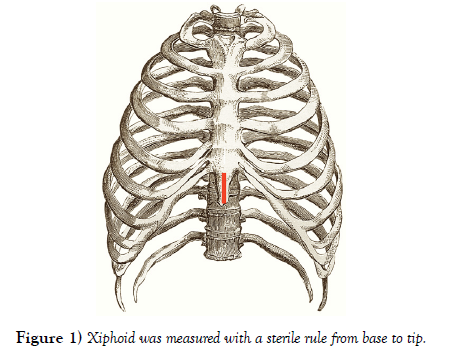
886 consecutive patients undergoing cardiac surgery via median sternotomy were entered into this study. A midline incision from the manubrio-sternal junction to the tip of the xiphoid was deepened to the periosteum. Inferiorly the aponeurotic attachments were divided to enable finger blunt dissection of the retro-sternal space. Prior to median sternotomy (which proceeded from the xiphoid upward using an air-powered jigsaw) the xiphoid was measured with a sterile rule from base to tip (Figure 1). Measurements were recorded to the closest 0.5cm. The xiphoid was considered vestigial when it measured 1cm or less. BSA was calculated from standard tables, based on the Du Bois formula [8].
The Independent samples t-test was used to compare means of continuous variables between two groups. The Chi squared test was used to investigate the association between categorical variables in different groups. Relationships between two continuous variables were assessed using the Pearson Correlation Test.
Results
There were 650 males and 236 females and mean age was 64.4 ± SD10.7 years (males 63.9 ± 10.0, females 65.9 ± 12.2, p=0.013). Mean age of females was significantly greater than that of males. Patient height (metres) was 1.63 ± 0.10 m (males 1.67 ± 0.08 m, females 1.54 ± 0.08 m p<0.001). BSA (metres2) was 1.83 ± 0.19 m2 (males 1.88 ± 0.17m2, females 1.67±0.17 m2 p<0.001). Xiphoid length (centimetres) was 2.77 ± 1.5 cm (males 3.27 ± 1.23 cm, females 1.37 ± 1.32 cm p<0.001). Mean height, BSA and xiphoid length of males was significantly greater than that of females.
There was a significant positive relationship between xiphoid length and body height in males (Pearson Correlation 0.094, p=0.016). There was no correlation between xiphoid length and body height in females (Pearson Correlation -0.028, p=0.668).
There was a significant positive relationship between xiphoid length and BSA in males (Pearson Correlation 0.107, p=0.006). There was no correlation between xiphoid length and BSA in females (Pearson Correlation 0.049, p=0.457).
The overall incidence of vestigial xiphoid was 110/887 or 12.41% with marked gender differences (males 19/650 or 2.93%, females 91/236 or 38.56% Chi squared=201.874, p< 0.001). The incidence of vestigial xiphoid was significantly higher in females.
Discussion
Body Surface Area rather than Body Mass Index was chosen as it more truly reflects body size [9]. Because of its numerous muscle attachments, we postulated that more robust abdominal muscles in a larger body might be associated with a longer xiphoid.
Allometry is the study dealing with the differential growth rates of parts of a living organism as well as shape differences in terms of ratios to the body dimensions. Isometric scaling describes the preservation of proportional relationships in different body sizes. In our study females exhibited negative allometry of the xiphoid when compared with males.
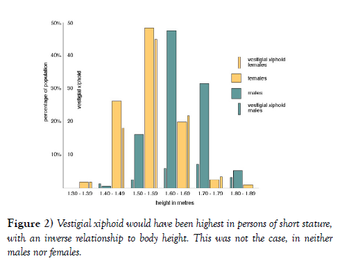
The correlation with body length and with BSA was strong in males but absent in females. Vestigial xiphoid was therefore not simply a short xiphoid in a diminutive body. Had this been the case, the incidence of vestigial xiphoid would have been highest in persons of short stature, with an inverse relationship to body height. This was not the case, in neither males nor females (Figure 2). The strong association of vestigial xiphoid (length ≤1 cm) with female gender suggests an alternative process.
Bone size and muscle mass and their correlation with handedness has been studied in adult upper limbs [10] as well as in fetuses [11] and the results suggest an inherited causation. The marked gender differences in our study also suggest an inherited basis.
Conclusion
Xiphoid length versus body height and body surface area exhibited a strong correlation in males, but not in females. The presence of vestigial xiphoid was significantly commoner in females, far outweighing the effect of body size.
REFERENCES
- Moore KL, Persaud TVN, Torchia MG. The developing human: clinically oriented embryology. 8th Ed. Elsevier Inc. Gurgaon, India. 2009;340.
- Mashriqi FD, Antoni AV, Tubbs RS. Xiphoid process variations: A review with an extremely unusual case report. Int J Anat Res. 2017; 9(8):1613.
- Yekeler E, Tunaci M, Dursun M, et al. Frequency of sternal variations and anomalies evaluated by MDCT. AJR Am J Roentgenol. 2006;186(4):956-960.
- Kumar NS, Bravian D, More AB. Xiphoid foramen and its clinical implications. Int Anat Res. 2014;2(2):340-343.
- HalvorsenTB, Anda SS, Naess AB, et al. Fatal cardiac tamponade after acupuncture through congenital sternal foramen.Lancet.1995; 1175.
- Shingleton A. Allometry: the study of biological scaling. Nature Education Knowledge 2010; 3(10): 2.
- Savage VM, Deeds EJ, Fontana F. Sizing up Allometric scaling theory. PLoS Computational Biology. 2008;9.
- Du Bois D, Du Bois EF. A formula to estimate the approximate surface area if height and weight be known. Arch Int Med. 1916;17(6):863-871.
- Sardinha LB, Silva AM, Minderico CS, et al. Effect of body surface area calculations on body fat estimates in non-obese and obese patients. Physiological Measurement 2006;27(11):1197-1209.
- Schell LM, Johnston FE, Smith DR, et al. Directional asymmetry of body dimensions among white adolescents. Am J Phys Anthorp. 1985;67:317- 322.
- Pande BS and Singh I. One-sided dominance in the upper limbs of human fetuses as evidenced by asymmetry in muscle and bone weight. J Anat. 1971;109:457-459.


