Bifid appendix: its embryologic explanation and clinical implications
Punita Sharma*, Anupama Mahajan, Poonam Verma and Anterpreet Arora
Department of Anatomy, Sri Guru Ram Das Institute of Medical Sciences and Research, Amritsar, India.
- *Corresponding Author:
- Punita Sharma
Professor,Department of Anatomy,Sri Guru Ram Das Institute of Medical Sciences and Research 242, Medical Enclave Circular Road, Amritsar,India
Tel: +91 183 2421508
E-mail: punitasalwan@gmail.com
Date of Received: January 20th, 2012
Date of Accepted: March 24th, 2012
Published Online: September 22nd, 2012
© Int J Anat Var (IJAV). 2012; 5: 38–40.
[ft_below_content] =>Keywords
appendix,bifid,classification,embryology,appendectomy
Introduction
The vermiform appendix present only in human beings, certain anthropoid apes and the wombat (a nocturnal, burrowing Australian marsupial) was probably first noted as early as the Egyptian civilization (3000 B.C.). During the mummification process, abdominal parts removed and placed in Coptic jars with inscriptions describing the contents as “worm of the intestines” were discovered [1].
The vermiform appendix is a narrow tube, which arises from the postero-medial caecal wall, approximately 2 cm below the end of the ileum, located in the right lower quadrant of abdomen. Its length varies from 2 to 20 cm, with an average of 9 cm [2].
Appendiceal variations are extremely rare: they have a reported incidence of between 0.004% and 0.009% of appendectomy specimens [3–5]. There have been some cases of true absence of the vermiform appendix reported, and average 100 cases of duplication have been reported to date [6].
Despite the notorious range of variations in characters and position natural to the human vermiform appendix, the extremes of variation –absence and duplication– affect this organ so very rarely that every authentic example of either is worthy of record [7]. Therefore, we thought of reporting this case of appendicular variation seen during routine cadaveric dissection.
Case Report
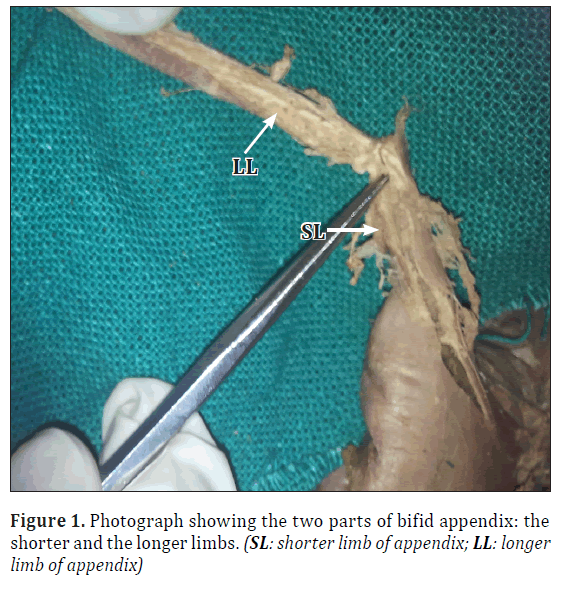
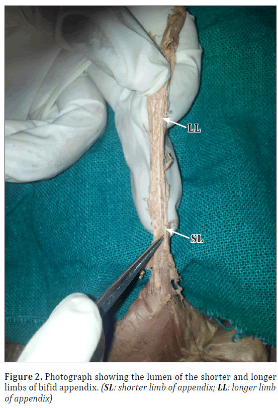
During routine dissection for undergraduate students in the Department of Anatomy at Sri Guru Ram Das Institute of Medical Sciences and Research, we came across an unusually formed bifid appendix. In our case, a 2.3 cm stump of appendix arose from the posteromedial part of a normal caecum, 2.5 cm below the ileo-caecal junction (Figures 1,2). Around 1.8 cm from the base of the stump of appendix, a 6.5 cm long limb arose with a slight change in direction. Blood supply to both came from the single appendicular artery arising from ileo-caecal artery lying in the single mesoappendix. On further exploration, the lumen was present in both the limbs. No other intra-abdominal malformations were present.
Discussion
Despite extraordinary advances in modern radiographic imaging and diagnostic laboratory investigations, the diagnosis of appendicitis remain essentially clinical requiring a mixture of observations, clinical acumen and surgical sense [8].
Congenital anatomical disorders of appendix are very rare. Therefore, it is important for several reasons that surgeons are aware of the potential anatomical variations and malposition of the vermiform appendix: first, a missed second appendix may result in serious clinical and medico-legal consequences; second, a double appendix can be confused with other intra-abdominal conditions; and finally, they can be associated with other congenital abnormalities [3].
More recently, on exploration, a perforated inflamed appendix was found coexisting with a second inflamed appendix, which was subserosal and retrocecal. Therefore, surgeons in emergency services should be aware of anatomical variations such as duplication and malposition of the appendix, even in patients with a history of previous appendectomy, because misdiagnosis of appendix duplication may lead to a poor clinical outcome and medicolegal issues [9].
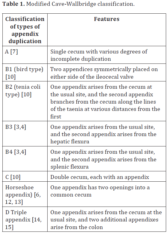
Appendix duplications were first classified by Cave in 1936 according to their anatomic location [7]. This classification system was updated and modified in 1963 by Wallbridge based on reported cases [10]. Again modified by Biermann in 1993 [11] (Table 1).
This system classifies three types of appendices: A, B, and C.
Embryology of the normal appendix has been defined. During the 5th fetal week it is the appendix which develops from a bud at the junction of the small and large bowel and undergoes rapid growth into a pouch. In the 6th week there is a transient nubbin surmounting the pouch indicative of being involved in the rapid development of the pouch, which is very strategically placed near the apex of the highly significant midgut loop. It is only after the fifth fetal month that the proximal end of this pouch, which has appeared to be a very insignificant structure up until this stage, starts growing differentially to give rise to the true caecum that continues to develop into infancy [16].
Although normal embryogenesis of appendix is known, but the pathogenesis of its duplication is unclear.
Furthermore, to explain the embryology of gastrointestinal duplications, following four theories put forward: The split notochord theory, failure of the normal regression of embryonic diverticula, the median septum formation, and partially twinning procedure [17]. Although the second theory may favor the pathogenesis, that there is failure of the normal regression of embryonic diverticula, none of them completely explains the embryology of appendix duplications. Nevertheless, Cave put forward two theories for the pathogenesis of duplex appendix: (a) supernumerary appendix due to persistence of a transient embryological structure; (b) appendiceal duplicity incidental to a more general affection of the primitive midgut [9]. Cave’s theories may explain this duplication, but they are not enough to explain all types.
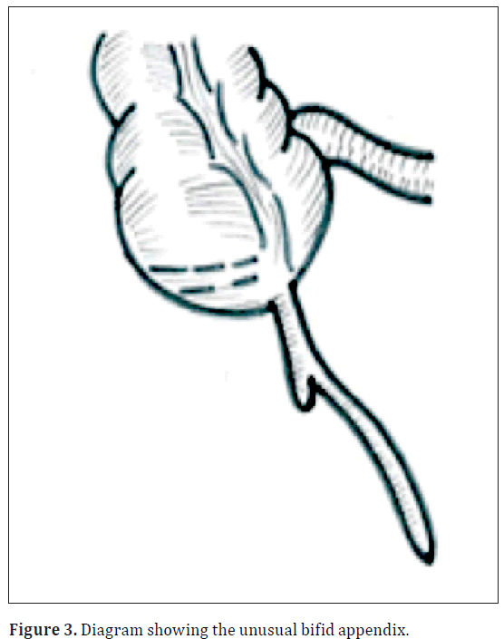
In our case (Figure 3), the long limb of appendix could be the supernumerary transient embryological structure, which has shifted to the normal growth of appendix (shorter limb) at some distance from the base. However, further investigation is required in the field of developmental anatomy.
Conclusion
Though considered to be a vestigial organ, the appendectomy is often the first major procedure performed by a surgeon in training. The variations and anomalies of appendix, though rare, can carry grave implications if missed during this surgery. Therefore, more such cases of appendicular variations should be reported so that surgeons in emergency services should be aware of unusual presentations such as duplication and malposition of the appendix.
Appendiceal duplication although rare and difficult to diagnose preoperatively, should be checked while operating for appendicular pathology in order to avoid serious clinical and medicolegal implications.
References
- Herrington JL Jr. The vermiform appendix: its surgical history. Contemp Surg. 1991; 39: 36–44.
- Standring S. Gray’s Anatomy: Anatomical Basis of Clinical Practice. 40th Ed., London, Elsevier. 2010: 1189–1190.
- Griffiths EA, Jagadeesan J, Fasih T, Mercer-Jones M. Bifid vermiform appendix: a case report. Curr Surg. 2006; 63: 176–178
- Kjossev KT, Losanoff JE. Duplicated vermiform appendix. Br J Surg. 1996; 83: 1259.
- Collins DC. A study of 50,000 specimens of the human vermiform appendix. Surg Gynecol Obstet. 1955; 101: 437–445.
- Calota F, Vasile I, Mogoanta S, Zavoi R, Pasalega M, Moraru E, Stoicea C. Horseshoe appendix: a extremely rare anomaly. Chirurgia (Bucur). 2010; 105: 271–274.
- Cave AJ. Appendix vermiformis duplex. J Anat. 1936; 70: 283–292.
- Bhasin SK, Khan AB, Kumar V, Sharma S, Saraf R. Vermiform appendix and acute appendicitis. JK Science. 2007; 9: 167–170.
- Canbay E, Akman E. Appendix perforation in appendix duplication in a man: a case report. J Med Case Reports. 2011; 5: 162.
- Wallbridge PH. Double appendix. Br J Surg. 1962; 50: 346–347
- Biermann R, Borsky D, Gogora M. Double appendicitis--a rare pathologic entity. Chirurg. 1993; 64: 1059–1061.
- Mesko TW, Lugo R, Breitholtz T. Horseshoe anomaly of the appendix: a previously undescribed entity. Surgery. 1989; 106: 563–566.
- DasGupta R, Reber PU, Patel AG. Horseshoe appendicitis. Eur J Surg. 1999; 165: 1095–1096.
- Tinckler LF. Triple appendix vermiformis: a unique case. Br J Surg. 1968; 55: 79–81.
- Uriev L, Maslovsky I, Monouskin Y, Ben-Dor D. Triple-barreled type of appendiceal triplication. Ann Diagn Pathol. 2006; 10: 160–161.
- Glover JW. The human vermiform appendix – a general surgeon’s reflections. EN Tech J. 1988; 3: 31–38.
- Jimenez SG, Oliver MR, Stokes KB, Morreau PN, Chow CW. Case report: Colonic duplication: a rare cause of obstruction. J Gastroenterol Hepatol. 1999; 14: 889–892.
Punita Sharma*, Anupama Mahajan, Poonam Verma and Anterpreet Arora
Department of Anatomy, Sri Guru Ram Das Institute of Medical Sciences and Research, Amritsar, India.
- *Corresponding Author:
- Punita Sharma
Professor,Department of Anatomy,Sri Guru Ram Das Institute of Medical Sciences and Research 242, Medical Enclave Circular Road, Amritsar,India
Tel: +91 183 2421508
E-mail: punitasalwan@gmail.com
Date of Received: January 20th, 2012
Date of Accepted: March 24th, 2012
Published Online: September 22nd, 2012
© Int J Anat Var (IJAV). 2012; 5: 38–40.
Abstract
Knowledge of vermiform appendix variations is important for the surgeons because diagnosis of appendicitis remain essentially clinical requiring a mixture of observations, clinical acumen and surgical sense in spite of extraordinary advances in modern radiographic imaging and diagnostic laboratory investigations. During routine dissection in the Department of Anatomy, we came across a bifid appendix, which does not fit into the routine classification of appendix duplication. A missed second appendix may result in serious clinical and medico-legal consequences and it can be confused with other intra-abdominal conditions.
-Keywords
appendix,bifid,classification,embryology,appendectomy
Introduction
The vermiform appendix present only in human beings, certain anthropoid apes and the wombat (a nocturnal, burrowing Australian marsupial) was probably first noted as early as the Egyptian civilization (3000 B.C.). During the mummification process, abdominal parts removed and placed in Coptic jars with inscriptions describing the contents as “worm of the intestines” were discovered [1].
The vermiform appendix is a narrow tube, which arises from the postero-medial caecal wall, approximately 2 cm below the end of the ileum, located in the right lower quadrant of abdomen. Its length varies from 2 to 20 cm, with an average of 9 cm [2].
Appendiceal variations are extremely rare: they have a reported incidence of between 0.004% and 0.009% of appendectomy specimens [3–5]. There have been some cases of true absence of the vermiform appendix reported, and average 100 cases of duplication have been reported to date [6].
Despite the notorious range of variations in characters and position natural to the human vermiform appendix, the extremes of variation –absence and duplication– affect this organ so very rarely that every authentic example of either is worthy of record [7]. Therefore, we thought of reporting this case of appendicular variation seen during routine cadaveric dissection.
Case Report
During routine dissection for undergraduate students in the Department of Anatomy at Sri Guru Ram Das Institute of Medical Sciences and Research, we came across an unusually formed bifid appendix. In our case, a 2.3 cm stump of appendix arose from the posteromedial part of a normal caecum, 2.5 cm below the ileo-caecal junction (Figures 1,2). Around 1.8 cm from the base of the stump of appendix, a 6.5 cm long limb arose with a slight change in direction. Blood supply to both came from the single appendicular artery arising from ileo-caecal artery lying in the single mesoappendix. On further exploration, the lumen was present in both the limbs. No other intra-abdominal malformations were present.
Discussion
Despite extraordinary advances in modern radiographic imaging and diagnostic laboratory investigations, the diagnosis of appendicitis remain essentially clinical requiring a mixture of observations, clinical acumen and surgical sense [8].
Congenital anatomical disorders of appendix are very rare. Therefore, it is important for several reasons that surgeons are aware of the potential anatomical variations and malposition of the vermiform appendix: first, a missed second appendix may result in serious clinical and medico-legal consequences; second, a double appendix can be confused with other intra-abdominal conditions; and finally, they can be associated with other congenital abnormalities [3].
More recently, on exploration, a perforated inflamed appendix was found coexisting with a second inflamed appendix, which was subserosal and retrocecal. Therefore, surgeons in emergency services should be aware of anatomical variations such as duplication and malposition of the appendix, even in patients with a history of previous appendectomy, because misdiagnosis of appendix duplication may lead to a poor clinical outcome and medicolegal issues [9].
Appendix duplications were first classified by Cave in 1936 according to their anatomic location [7]. This classification system was updated and modified in 1963 by Wallbridge based on reported cases [10]. Again modified by Biermann in 1993 [11] (Table 1).
This system classifies three types of appendices: A, B, and C.
Embryology of the normal appendix has been defined. During the 5th fetal week it is the appendix which develops from a bud at the junction of the small and large bowel and undergoes rapid growth into a pouch. In the 6th week there is a transient nubbin surmounting the pouch indicative of being involved in the rapid development of the pouch, which is very strategically placed near the apex of the highly significant midgut loop. It is only after the fifth fetal month that the proximal end of this pouch, which has appeared to be a very insignificant structure up until this stage, starts growing differentially to give rise to the true caecum that continues to develop into infancy [16].
Although normal embryogenesis of appendix is known, but the pathogenesis of its duplication is unclear.
Furthermore, to explain the embryology of gastrointestinal duplications, following four theories put forward: The split notochord theory, failure of the normal regression of embryonic diverticula, the median septum formation, and partially twinning procedure [17]. Although the second theory may favor the pathogenesis, that there is failure of the normal regression of embryonic diverticula, none of them completely explains the embryology of appendix duplications. Nevertheless, Cave put forward two theories for the pathogenesis of duplex appendix: (a) supernumerary appendix due to persistence of a transient embryological structure; (b) appendiceal duplicity incidental to a more general affection of the primitive midgut [9]. Cave’s theories may explain this duplication, but they are not enough to explain all types.
In our case (Figure 3), the long limb of appendix could be the supernumerary transient embryological structure, which has shifted to the normal growth of appendix (shorter limb) at some distance from the base. However, further investigation is required in the field of developmental anatomy.
Conclusion
Though considered to be a vestigial organ, the appendectomy is often the first major procedure performed by a surgeon in training. The variations and anomalies of appendix, though rare, can carry grave implications if missed during this surgery. Therefore, more such cases of appendicular variations should be reported so that surgeons in emergency services should be aware of unusual presentations such as duplication and malposition of the appendix.
Appendiceal duplication although rare and difficult to diagnose preoperatively, should be checked while operating for appendicular pathology in order to avoid serious clinical and medicolegal implications.
References
- Herrington JL Jr. The vermiform appendix: its surgical history. Contemp Surg. 1991; 39: 36–44.
- Standring S. Gray’s Anatomy: Anatomical Basis of Clinical Practice. 40th Ed., London, Elsevier. 2010: 1189–1190.
- Griffiths EA, Jagadeesan J, Fasih T, Mercer-Jones M. Bifid vermiform appendix: a case report. Curr Surg. 2006; 63: 176–178
- Kjossev KT, Losanoff JE. Duplicated vermiform appendix. Br J Surg. 1996; 83: 1259.
- Collins DC. A study of 50,000 specimens of the human vermiform appendix. Surg Gynecol Obstet. 1955; 101: 437–445.
- Calota F, Vasile I, Mogoanta S, Zavoi R, Pasalega M, Moraru E, Stoicea C. Horseshoe appendix: a extremely rare anomaly. Chirurgia (Bucur). 2010; 105: 271–274.
- Cave AJ. Appendix vermiformis duplex. J Anat. 1936; 70: 283–292.
- Bhasin SK, Khan AB, Kumar V, Sharma S, Saraf R. Vermiform appendix and acute appendicitis. JK Science. 2007; 9: 167–170.
- Canbay E, Akman E. Appendix perforation in appendix duplication in a man: a case report. J Med Case Reports. 2011; 5: 162.
- Wallbridge PH. Double appendix. Br J Surg. 1962; 50: 346–347
- Biermann R, Borsky D, Gogora M. Double appendicitis--a rare pathologic entity. Chirurg. 1993; 64: 1059–1061.
- Mesko TW, Lugo R, Breitholtz T. Horseshoe anomaly of the appendix: a previously undescribed entity. Surgery. 1989; 106: 563–566.
- DasGupta R, Reber PU, Patel AG. Horseshoe appendicitis. Eur J Surg. 1999; 165: 1095–1096.
- Tinckler LF. Triple appendix vermiformis: a unique case. Br J Surg. 1968; 55: 79–81.
- Uriev L, Maslovsky I, Monouskin Y, Ben-Dor D. Triple-barreled type of appendiceal triplication. Ann Diagn Pathol. 2006; 10: 160–161.
- Glover JW. The human vermiform appendix – a general surgeon’s reflections. EN Tech J. 1988; 3: 31–38.
- Jimenez SG, Oliver MR, Stokes KB, Morreau PN, Chow CW. Case report: Colonic duplication: a rare cause of obstruction. J Gastroenterol Hepatol. 1999; 14: 889–892.


