Case Report of Bilateral Supernumerary Renal Vessels
Received: 24-Jun-2020 Accepted Date: Aug 10, 2020; Published: 17-Aug-2020, DOI: 10.37532/1308-4038.20.13.9
This open-access article is distributed under the terms of the Creative Commons Attribution Non-Commercial License (CC BY-NC) (http://creativecommons.org/licenses/by-nc/4.0/), which permits reuse, distribution and reproduction of the article, provided that the original work is properly cited and the reuse is restricted to noncommercial purposes. For commercial reuse, contact reprints@pulsus.com
Abstract
Renal arteries, in most of the cases, arise from the abdominal aorta, at the level of the second lumbar vertebra. At the same level, renal veins join the inferior vena cava. However, due to the intricate development of the kidneys, the alterations of these vessels in reference to their origin, position, pattern and number occur fairly frequently. Knowing the possible variations of blood supply to the kidneys is a necessity for doing any renal surgery including transplantation, surgery on aortic aneurysm or urologic malformations. We report multiple variations in the renal vessels found during routine dissection of a cadaver of a 90 years old man. There were four renal arteries and veins on the left side, and one supernumerary renal artery on the right side. In addition, the hilum on the left side was shifted ventrally, and a considerable portion of the renal pelvis was excluded from the renal sinus.
Keywords
Accessory renal vessel; Renal surgery; Renal transplantation; Variation
Introduction
The development of the kidney and its vessels is a fairly intricate process and – accordingly – their anatomy and blood supply are very complex. Due to the delicacy of the development, several malformations may occur involving both structure and circulation of the kidney or of the urinary tract. Anatomical variations (not necessarily resulting in pathological consequences) mostly affect the renal arteries. Knowing the alterations of these vessels is of equally high importance both in renal surgery and renal transplantation owing to their relatively high prevalence in the population. Vascular anomalies were found in about 30% of cadavers aquired for transplantation purposes or in patients who had CT angiography [1].
The kidneys develop from the metanephros in the caudal (pelvic) region, later shift to more cranial position in the retroperitoneum. In the pelvis, the metanephros receives its arterial supply from the lateral visceral segmental branches of the aorta. During its ascent, the kidney is supplied by continuously developing segmental arteries that originate from gradually higher and higher levels of the abdominal aorta. Meanwhile the lower arteries degenerate, but occasionally some may persist resulting in vascular variations.
These variants are described as accessory or aberrant renal arteries. Accessory renal arteries are additional arteries that pass along with normal renal arteries through the hilum. In contrast, aberrant arteries are those which enter the kidney by piercing the renal parenchyme, either above (upper polar artery) or below (lower polar artery) the hilum [2].
The classification of the accessory renal arteries is based on their origin:
(1) supernumerary renal arteries arising from the abdominal aorta, or
(2) from the main renal artery, or
(3) from other arteries.
According to the literature, single renal arteries were observed in 73% of cases, 48% in male and 52% in female [3]. The accessory renal artery may occur in 30-35% of the population [1], usually on the left side [4] and 18% of them originates from the abdominal aorta. 12% of accessory renal arteries come from the renal artery. The incidence of bilateral variations is 10%, those of unilateral ones is 21% [5].
The well-known congenital renal anomaly is the horseshoe kidney found in 0,25% of the general population [6], and is twice as frequent in men as in women [7]. A wide variety of associated anomalies coexists with horseshoe kidney, arterial anomalies are quite common, the occurence of them is 60- 80% [6].
More than one accessory renal arteries were observed in 6-8% of cases. Four renal arteries on the same side were sparse (0,4-0,5%). Very rarely the accessory renal arteries originated from the primary or secundary branches of the abdominal aorta, i.e. testicular artery, median sacral artery, ovarian artery, celiac trunk, right colic artery, superior suprarenal artery, inferior phrenic artery, splenic artery, thoracic aorta or contralateral renal artery [4,8].
Okamoto et al. [9] grouped the inferior supernumerary renal arteries into three classes:
(1) artery of lower origin that passes in front of the inferior vena cava and behind the ureter,
(2) artery of middle origin passing in front of both the inferior vena cava and the ureter,
(3) artery of upper origin that passes behind the inferior vena cava and in front of the ureter or renal pelvis.
In addition, there was a tendency for the lower origin type to have a ureteric branch, while the middle and upper origin types frequently had a gonadal branch.
Materials and Methods
This study was based on dissection of an elderly male cadaver. During routine abdominal dissection for medical undergraduates, the kidneys along with their arteries were disclosed and the morphological variations of renal vessels were noted. During the course of dissection, most abdominal viscera were removed and the abdominal vessels (abdominal aorta, inferior vena cava) cleaned. The kidneys with the supernumerary renal vessels were preserved as specimens for teaching purposes. Pictures showing the cleaned structures and the retroperitoneal topography were taken.
Case Report
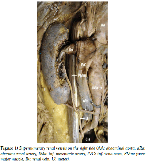
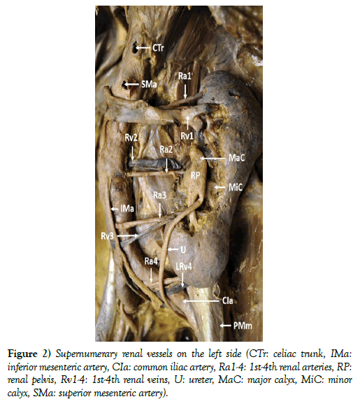
In our case (male aged 90, with no renal disease in the case history), we found one supernumerary renal artery on the right side (Figure 1). On the left side (Figure 2), there were four renal arteries, the highest representing the normal anatomical position. Each artery was accompanied by the corresponding renal vein. Moreover, the left hilum was shifted to the ventral surface of the kidney, and quite a big portion of the renal pelvis was excluded from the renal sinus. Since the persistent renal arteries interfered with the upward migration of the kidneys, both kidneys had markedly elongated shape, and their lower poles were extending more inferiorly than usual.
The sizes of the right kidney were 13,5 cm longitudinally, 5,3 cm transversely at the widest portion, and 4,5 cm antero-posteriorly. The corresponding sizes of the left kidney were 15,5 cm longitudinally, 5,5-6 cm transversely at both poles and 4 cm at the hilum, and 5,5 cm antero-posteriorly.
Detailed morphology of the accessory renal vessels
Right side (Figure 1)
The single aberrant renal artery (aRa) originated from the very beginning of the right common iliac artery (CIa), and entered the kidney near to its inferior pole. There was no accessory renal vein on this side.
Left side (Figure 2)
The upper three renal arteries originated from the abdominal aorta (AA). The highest (Ra1) emerged from the aorta on its left side, halfway between superior (SMa) and inferior mesenteric arteries (IMa). It entered the parenchyme near the superior pole of the kidney. The second artery (Ra2) also originated from the left side of the aorta, but lower, at the level of inferior mesenteric artery, and passed through the hilum. The third (Ra3) was leaving the the abdominal aorta on its anterior surface slightly to the right of the midline and 2 cm above the aortic bifurcation. It reached the kidney at the inferior end of its hilum. The fourth artery (Ra4) was a branch of the left common iliac artery (CIa) arising from its medial side. After passing anterior to the common iliac artery, it entered the parenchyme at the inferior pole.
The left renal veins followed the corresponding arteries. The highest vein (Rv1) turned in front of the abdominal aorta before entering the inferior vena cava. The second (Rv2) also joined the inferior vena cava, but after passing behind the abdominal aorta. The third vein (Rv3) crossed the abdominal aorta on its anterior side and joined the inferior vena cava on its right side. The lowest vein (Rv4) opened into the left common iliac vein on its lateral side.
The morphology of the renal pelves
Right side (Figure 1)
In spite of the enlarged hilum, its structures were arranged in the normal anatomical sequence.
Left side (Figure 2)
Together with the altered hilum, the renal pelvis (RP) was also shifted anteriorly. Normally, the renal pelvis is situated in the renal sinus. In our case, the entire pelvis and the last portions of major calices (MaC) were located outside the renal hilum, i.e. free of renal parenchyme coverage. The ureter (U) turned medially in front of the lower major calyx and continued down to the lesser pelvis anterior to the lowest, aberrant renal vessels (Ra4, Rv4).
Discussion
According to classical anatomy, the kidney is supplied by a single renal artery originating from abdominal aorta, but in many cases, variations of vascularization exist. The various types of renal arteries (accessory, additional, supplementary, and aberrant), their position, way of their entry to the kidney, and segmentation were studied extensively by a number of authors, but the generally accepted and precise terminology for these arteries has not been unified by the majority of them. To minimalize complications caused by unexpected bleeding, knowledge of the variations of the origin and branching pattern of renal vessels is of surpassing value to surgeons performing any operation on kidneys (transplantation, vascular reconstruction, partial or total resection, etc).
The continously developing, more and more sophisticated surgical methods necessitate the knowledge of more and more minute detailes of classical and surgical anatomy. Many minimal invasive surgical procedures, e.g. the lateral intrabody fusion performed in the retroperitoneum or on the thoracolumbal part of the vertebral column are carried out through the iliopsoas muscle. This approach rizes the possibility of injury of not only the normal, but - with even higher probability – the incidental supranumerary arteries. Variations in the pattern of renal vascularization may lead to circulatory disorders. Aneurysm may also develop on supernumerary renal artery [10]. The variations in the renal vascularization are either recognized and described in the clinial practice if they produce significant symptoms using ultrasound, CT, MR, or during the dissections.
Conclusion
In our case we found a rare variant of vascular supply of the kidneys. There were four supernumerary renal arteries accompanied by the corresponding veins on the left side, and one on the right. Moreover, the hilum on the left side was shifted ventrally. In spite of these anatomical variation, the 90 years old man was free of any symptoms.
REFERENCES
- Pollak R, Prusak BF, Mozes MF. Anatomic abnormalities of cadaver kidneys procured for purposes of transplantation. Am Surg. 1986;52:233-5.
- Merklin RJ, Michels NA. The variant renal and suprarenal blood supply with data on the inferior phrenic, ureteral and gonadal arteries: a statistical analyzis based on 185 dissections and review of the literature. J Intnl Coll Surgeons. 1982;29:41-76.
- Staśkiewicz G, Jajko K, Torres K, et al. Supernumerary renal vessels: analysis of frequency and configuration in 996 computed tomography studies. Fol Morphol. 2016;75:245-50.
- Mao QH, Li J. An accessory renal artery originating from the testicular artery, a rare variant. Indian J Surg. 2015;77:549-50.
- Anuoma G, Raman G, Rikki S. Congenital variations of renal veins: embryological background and clinical implications. J Clin Diagn Res. 2011;6:1140-3.
- Ichikawa T, Tanno K, Okochi T, et al. Evaluation of renal artery anomalies associated with horseshoe kidney using CT angiography. Tokai J Exp Clin Med. 2015;40:16-21.
- Bauer SB, Perlmutter AD, Retik AB. Anomalies of the upper urinary tract. In: Walsh PC, editor. Campbell’s Urology 6th ed. Philadelphia WB Saunders. 1992;1357-44.
- Bojisen E. Angiographic studies of the anatomy of single and mulitple renal arteries. Acta Radiol. Suppl. 1959;183:7.
- Okamoto K, Kodama K, Kawai K, et al. The inferior supernumerary renal arteries: a classification into three types. Ann Anat. 2006;188:49-53.
- Nishie R, Toya N, Fukushima S, et al. Prophylactic accessory renal artery coil embolization for prevention of type II endoleak following endovascular aneurysm repair: a case report. Surg Case Rep. 2017;3:58.


