Case Report on Variations in the Branching Pattern of the Brachial Artery: Clinical Significance and Surgical Implications
Received: 04-Jun-2023, Manuscript No. ijav-23-6531; Editor assigned: 05-Jun-2023, Pre QC No. ijav-23-6531 (PQ); Accepted Date: Jun 23, 2023; Reviewed: 19-Jun-2023 QC No. ijav-23-6531; Revised: 23-Jun-2023, Manuscript No. ijav-23-6531 (R); Published: 30-Jun-2023, DOI: 10.37532/1308-4038.16(6).273
Citation: Hvizdosova N. Case Report on Variations in the Branching Pattern of the Brachial Artery: Clinical Significance and Surgical Implications. Int J Anat Var. 2023;16(6):321-322.
This open-access article is distributed under the terms of the Creative Commons Attribution Non-Commercial License (CC BY-NC) (http://creativecommons.org/licenses/by-nc/4.0/), which permits reuse, distribution and reproduction of the article, provided that the original work is properly cited and the reuse is restricted to noncommercial purposes. For commercial reuse, contact reprints@pulsus.com
Abstract
The brachial artery is a major vessel of the upper limb, supplying oxygenated blood to the muscles and tissues. Variations in its branching pattern are relatively common and have important clinical implications for surgical procedures, interventional radiology, and diagnostic imaging. In this case report, we present a unique anatomical variation in the branching pattern of the brachial artery observed during a routine cadaveric dissection. The arterial variation involved an additional branch arising from the brachial artery, which can potentially impact the accuracy of clinical procedures and treatment decisions. Understanding such variations is crucial for healthcare professionals to ensure patient safety and optimize clinical outcomes.
Keywords
Brachial artery; Anatomical variation; Branching pattern; Clinical anatomy; Surgical implications
INTRODUCTION
The brachial artery is a continuation of the axillary artery, extending from the lower border of the teres major muscle to the cubital fossa in the anterior aspect of the elbow. It is a critical vascular structure supplying the upper limb, and its branching pattern has been well-described in anatomical literature. However, variations in the branching pattern are not uncommon and can have significant clinical implications [1].
Variations in the branching pattern of the brachial artery have been attributed to embryological development, genetic factors, and evolutionary changes. These variations may involve the absence, duplication, or aberrant course of branches, leading to variations in the blood supply to the forearm, hand, and associated muscles [2]. Awareness of these variations is of paramount importance to surgeons, radiologists, and clinicians involved in various interventions and procedures of the upper limb.
In this case report, we present a unique anatomical variation in the branching pattern of the brachial artery identified during a routine cadaveric dissection. The case demonstrates the importance of understanding and recognizing such variations to ensure safe and effective clinical management [3-4].
CASE REPORT
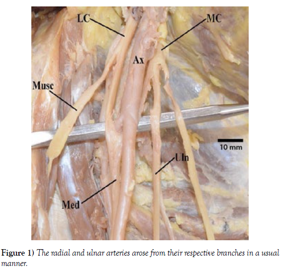
During a routine cadaveric dissection of the upper limb, a previously unreported variation in the branching pattern of the brachial artery was observed. In the present case, the brachial artery followed its normal course from the axilla to the cubital fossa, but at the level of the mid-arm, an additional branch originated from its lateral aspect. This branch, which was named the “accessory brachial artery” (ABA), coursed parallel to the main brachial artery for a short distance before dividing into two branches in the forearm. The radial and ulnar arteries arose from their respective branches in a usual manner.’
The presence of the ABA could have significant clinical implications in various medical and surgical scenarios. For instance, during arterial cannulation for invasive blood pressure monitoring or intravenous drug administration, the presence of the ABA may increase the risk of inadvertent puncture, leading to potential complications such as hematoma formation or distal ischemia. Similarly, in arterial reconstruction procedures or trauma management, surgeons should be aware of such anatomical variations to avoid inadvertent injury to the accessory vessel or compromise the blood supply to the forearm and hand (Figure 1).
DISCUSSION
The variation observed in this case, involving an accessory branch arising from the brachial artery has not been previously reported in the literature. Variations in the branching pattern of the brachial artery are relatively common, with reported prevalence rates ranging from 13% to 30% in various anatomical studies. However, most commonly reported variations involve the absence or duplication of branches rather than the presence of an accessory vessel [5].
Understanding the anatomical variations of the brachial artery is crucial for several clinical specialties. Surgeons performing vascular reconstruction procedures, such as arterial bypass grafting or angioplasty, need to be aware of the potential presence of accessory vessels to prevent inadvertent damage during surgery. Interventional radiologists performing procedures like transradial catheterization or angiography should also be cautious to avoid complications associated with these variations [6-8].
In addition to surgical implications, knowledge of the anatomical variations in the branching pattern of the brachial artery is essential for accurate diagnostic imaging and interpretation. Radiologists and clinicians interpreting computed tomography angiography (CTA), magnetic resonance angiography (MRA), or Doppler ultrasound studies of the upper limb should be familiar with these variations to avoid misinterpretation of images or diagnostic errors.
Moreover, anatomical variations such as the presence of an accessory branch in the brachial artery may affect the interpretation of pulse examination in clinical practice. Pulse assessment in the radial and ulnar arteries is a routine examination technique to evaluate peripheral arterial circulation. The presence of an accessory vessel can result in an unusual or absent pulse, potentially leading to diagnostic confusion or inappropriate clinical management [9-10].
CONCLUSION
This case report highlights a unique anatomical variation in the branching pattern of the brachial artery, involving the presence of an accessory branch. Variations in the brachial artery’s branching pattern are not uncommon and can have significant clinical implications for surgical procedures, diagnostic imaging, and clinical decision-making. Healthcare professionals involved in the management of upper limb pathologies should be aware of these variations to ensure safe and effective care. Further research is warranted to investigate the prevalence, clinical significance, and potential complications associated with these variations.
ACKNOWLEDGEMENT
The authors would like to acknowledge the cadaveric donor and their family for their valuable contribution to medical education and research.
CONFLICT OF INTEREST
The authors declare no conflicts of interest related to this case report.
REFERENCES
- Veltman CE, van der Hoeven BL, Hoogslag GE, Boden H, Kharbanda RK, et al. Influence of coronary artery dominance on short- and log-term outcomes in patients after ST-segment elevation myocardial infarction. Eur Heart J. 2015; 36:1023-1030.
- Galiuto L. How to access functional significance of myocardial bridging in athletes: a personalized medicine approach. Biomed J Sci & Tech Res. 2020; 26(2): 004-313.
- Navarro A, Sladden D, Casha A, Manche A. The difficulty in identifying and grafting an intramuscular coronary artery. Malta Med J. 2019; 3(1):14-16.
- Ibarrola M. Myocardial bridge a forgotten condition: A review. Clin Med Img Lib. 2021; 7:182.
- Jiang L, Zhang M, Zhang H, Shen L, Shao L, et al. A potential protective element of myocardial bridge against severe obstructive atherosclerosis in the whole coronary system. BMC Cardiovasc Disord. 2018; 18(1):105.
- Aricatt DP, Prabhu A, Avadhani R, Subramanyam K, Ezhilan J, et al. A study of coronary artery dominance and its clinical significance. Folia Morphol. 2023; 82(1): 102-107.
- Abu-Assi E, Castineira-Busto M, Gonzalez-Salvado V, Raposeiras-Roubin S, Abumuaileq RR-Y, et al. Coronary artery dominance and long-term prognosis in patients with ST-segment elevation myocardial infarction treated with primary angioplasty. Rev Esp Cardiol. 2016; 69(1):19-27.
- Vural A, Cicek ED. Is asymmetry between vertebral arteries related to cerebral dominance? Turk J Med Sci. 2019; 49: 1721-1726.
- Rusu MC, Vrapclu AD, Lazar M. A rare variant of accessory cerebral artery. Surg Radiol Anat. 2023; 45(5):523-526.
- Mani K. Absent internal carotid artery in the circle of Willis. IOSR-JDMS. 2015; 14(11): 38-40.
Indexed at, Google Scholar, Crossref
Indexed at, Google Scholar, Crossref
Indexed at, Google Scholar, Crossref
Indexed at, Google Scholar, Crossref
Indexed at, Google Scholar, Crossref
Indexed at, Google Scholar, Crossref
Indexed at, Google Scholar, Crossref
Indexed at, Google Scholar, Crossref


