Common celio-mesentric renal trunk: An extremely rare anatomic variant
Received: 01-Apr-2018 Accepted Date: Apr 16, 2018; Published: 30-Apr-2018, DOI: 10.37532/1308-4038.18.11.55
Citation: Shaw M, Rajagopal R, Jagia P, et al. Common celio-mesentric renal trunk: An extremely rare anatomic variant. Int J Anat Var. 2018;11(2):55-58.
This open-access article is distributed under the terms of the Creative Commons Attribution Non-Commercial License (CC BY-NC) (http://creativecommons.org/licenses/by-nc/4.0/), which permits reuse, distribution and reproduction of the article, provided that the original work is properly cited and the reuse is restricted to noncommercial purposes. For commercial reuse, contact reprints@pulsus.com
Abstract
Anatomic variations in the branches of abdominal aorta are very common. Among them, the most common variations are those involving the renal arteries. We report the case of a 9 year old boy with complaints of intermittent abdominal pain, bilateral lower limb claudication & hypertension that was referred for evaluation. An extremely rare variation in the origins of visceral and renal arteries was noted in this patient with the abdominal aorta giving rise to a common trunk at T12-L1 level which further branched into celiac artery, superior mesenteric artery and both renal arteries. The native aorta distal to the origin of this common trunk showed circumferential wall thickening with decreased luminal diameter suggestive of aorto-arteritis.
Keywords
Celio-mesentric renal trunk; Angiography; Mesentric vessels; Renal arteries
Introduction
Variations in morphology of the abdominal aorta and its branches are of interest to the vascular physician, surgeon and interventional radiologist. This is because the geometry of vessels and branches determine the flow dynamics and help in deciding the appropriate surgical or interventional approach. The involvement of different branches also gives clues about various pathological processes which have specific patterns of vessel involvement. The most common variant anatomy of the celiac axis is the common origin of hepatic, splenic and left gastric artery from the abdominal aorta (AA) [1]. Similarly, the most common variant anatomy of SMA is right hepatic artery arising from superior mesenteric artery (SMA), instead of the common hepatic artery, also called as replaced right hepatic artery. Accessory renal arteries are the most common variation of renal arteries [1-8]. Variations and clinical significance of the renal artery origin, course and branching pattern have been frequently described in literature, which are of importance during endovascular and surgical procedures [8].
This case report demonstrates an extremely rare variation in branching of abdominal aorta where celiac axis, SMA and both renal arteries are seen to arise from a common celio-mesentric renal trunk (CMRT), arising from the abdominal aorta at the T12-L1 level.
Case Report
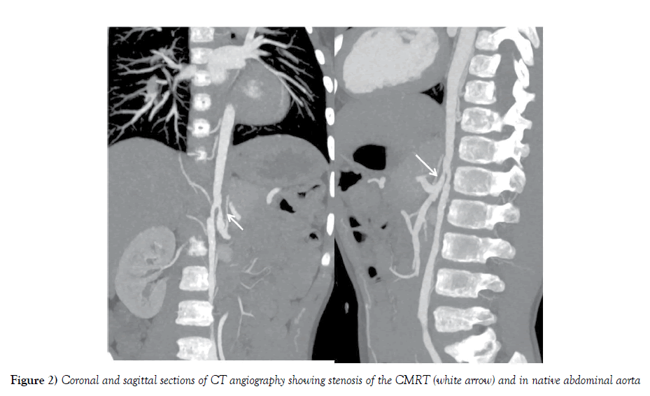
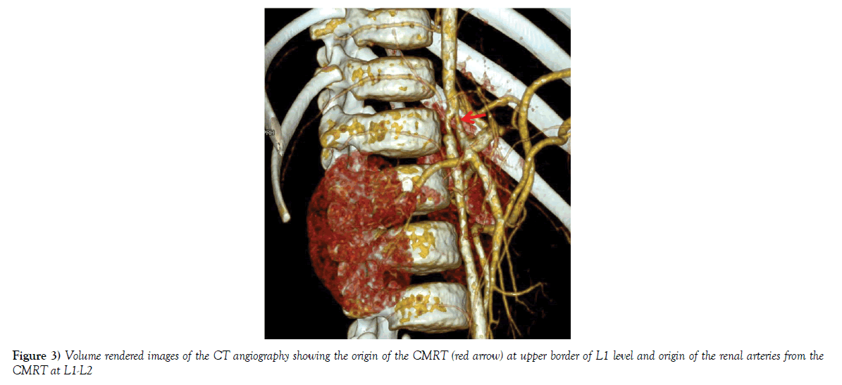
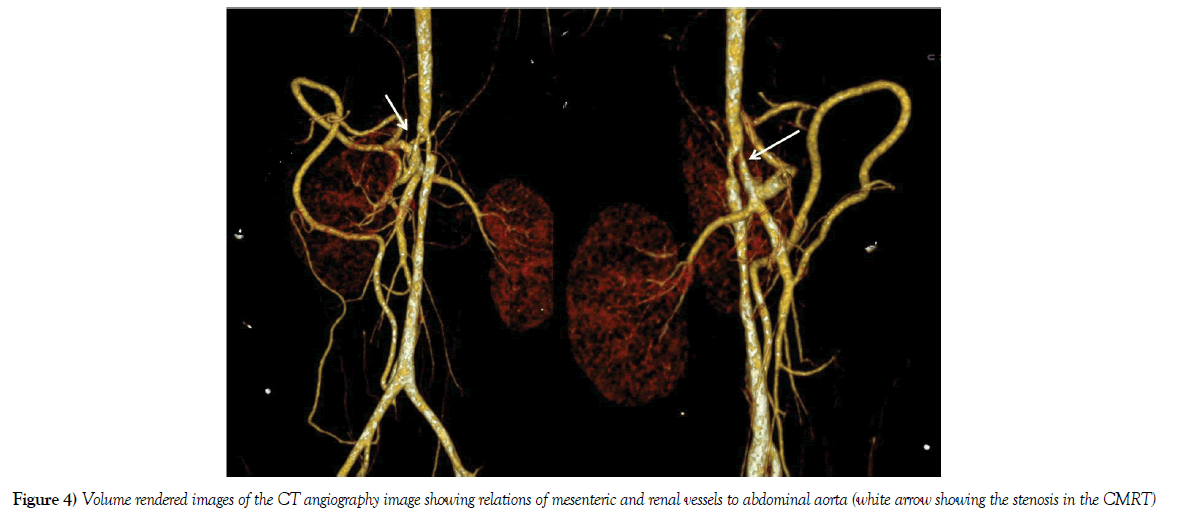
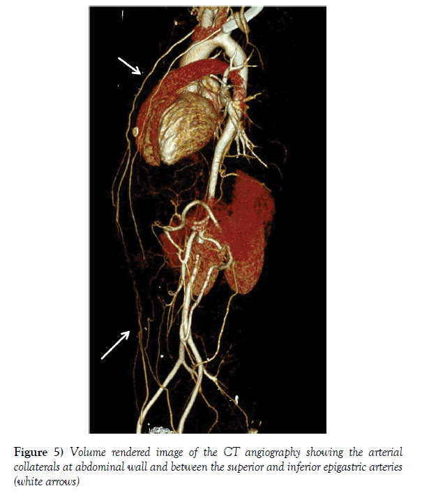
A nine year old boy was referred to our department of Cardiovascular Radiology with complaints of intermittent abdominal pain, lower limb fatigue and hypertension for evaluation. Clinical examination revealed feeble bilateral lower limb pulses. His blood pressure was 154/90 mmHg in left upper limb and 110/80 in lower limb. Echocardiography showed mild left ventricular systolic dysfunction. Complete blood counts and renal function tests were normal. A frontal chest radiograph of the child showed mild cardiomegaly. Based on a clinical suspicion of non-specific aortoarteritis, he underwent a Computed Tomography angiography (CTA) of aorta in a 384 slice dual source CT scanner (Somatom Force, Siemens, Germany) with intravenous administration of 30 ml non-ionic iodinated contrast (Iohexol, Omnipaque 350 mgI/ml, GE) using the bolus tracking technique with region of interest (ROI) placed in upper descending thoracic aorta (DTA). The ascending aorta, aortic arch, DTA along with the aortic arch branches was normal. The abdominal aorta had a single anterior branch - the common celio-mesentric renal trunk (CMRT) which was giving rise to celiac axis, SMA and both renal arteries (Figures 1 and 2). The native abdominal aorta distal to this branch showed diffuse circumferential wall thickening causing upto 50% luminal stenosis (Figure 3). The anterior CMRT had following branches: 1) Celiac axis dividing to left gastric artery, hepatic artery, splenic artery and bilateral inferior phrenic artery; 2) superior mesenteric artery and 3) bilateral renal arteries (Figure 4). The ostio-proximal segment of CMRT showed upto 50% stenosis with mild post stenotic dilatation before branching. The termination of the common trunk was by division into smaller branches supplying the small intestine (representing the branches of SMA). Multiple collaterals were seen in the anterior abdominal wall (Figure 5). The blood pressure was under control by optimum medical management till the latest follow up and thus he was maintained on medical therapy with graded exercise training.
Discussion
Different anatomical variations of the branches of the abdominal aorta namely, the mesenteric and renal vasculature have been previously described in literature (Table 1). Inferior mesenteric artery, which typically originates from abdominal aorta at the level of L3 vertebra, has little variation of its origin [8]. Origin of renal arteries from mesenteric vessels has been described only in few isolated case reports to the best of our knowledge (Table 2). This case demonstrates the common celio-mesentric renal trunk which is an extremely rare variation of the abdominal aortic branching pattern, previously described only in a 14 year old female child by Connolly et al. [4].
| Sl.no | Variation and description | Incidence |
|---|---|---|
| A. Celiac axis | ||
| 1. | Common trunk with bifurcation to hepato-splenic trunk and left gastric artery | 50-76% |
| 2. | Direct origin of common hepatic artery, splenic artery and left gastric artery | 10-19% |
| 3. | Quadrifurcation/ pentafurcation- Dorsal pancreatic artery, gastro duodenal artery and right/left hepatic artery along with splenic and left gastric artery from celiac axis | Upto 10% |
| 4. | Common hepato splenic trunk and left gastric artery from aorta | 1.5-4.4% |
| 5. | Hepato mesentric and gastro splenic trunk | 0.5-2.6% |
| 6. | Common celio-mesentric trunk | 1.1% |
| B. SMA variation: | ||
| 7. | Normal anatomy of SMA | ~68% |
| 8. | Middle colic-right colic trunk | Upto 52% |
| 9. | Accessory right colic artery | 8-10% |
| 10. | Common origin of superior mesenteric and hepatic arteries | 0.5% |
| C. Renal artery variation | ||
| 11. | Typical bilateral single renal artery | 72.6% |
| 12. | Early branching of renal artery | 6.7% |
| 13. | Additional hilar artery | 7.0% |
| 14. | Superior renal polar artery | 4.7% |
| 15. | Inferior renal polar artery | 8.7% |
| 16. | Inferior and superior renal polar arteries | 0.3% |
Table 1: Variations of Celiac, superior mesenteric and renal arteries [2,7-12]
| Sl.no. | Authors | Evaluating technique | Description of anatomic variation |
|---|---|---|---|
| 1. | Connolly et al. [3] | CTA and Angiography | Common CMRT giving rise to celiac, SMA and both renal arteries |
| 2. | Bamak et al. [6] | Autopsy | Right renal artery as a branch from SMA |
| 3. | Dalcik et al. [13] | Autopsy | Common trunk giving rise to SMA and right renal artery |
| 4. | Lacout et al. [14] | CTA | Right renal artery origin from SMA |
| 5. | Daraghmeh et al. [15] | CTA | Celiomesentric trunk gives rise to celiac, SMA and right renal artery; left renal artery arises from aorta |
| 6. | Bartoli et al. [16] | CTA and Angiography | Celiomesentric trunk gives rise to celiac, SMA and left renal artery; right renal artery arises from aorta |
Table 2: Various variations of the branching (common origin) pattern of the mesenteric and renal arteries:
Embryologically, the celiac axis is the vessel of the foregut and SMA is the artery of the mid gut. A common trunk giving rise to both celiac and SMA can be explained by persistence of ventral longitudinal anastomosis cranial to the fourth root of ventral branches of abdominal aorta with regression of either first or fourth ventral root causing a common celio-mesentric trunk [3,9-14]. The common trunk giving rise to celiac, SMA and both renal arteries is an extremely rare variant, the embryological basis of which is unknown, although Bamak et al. have suggested that during embryological development and ascent of renal tissues, the renal tissues may recruit a branch from SMA which may later on persist as the main renal artery [2]. In the patient described by Conolly et al., the common aortic trunk was seen giving off the left renal artery from its proximal part of common trunk; followed by celiac artery & SMA and right renal artery was originating between celiac and SMA [4]. Our patient also had two segments of stenoses, one at the native abdominal aorta after the origin of common CMRT & another at the ostio-proximal part of CMRT itself, due to involvement by vasculitis. This anatomic variation is of extreme importance, as selective catheterization of the visceral arteries during endovascular procedures may be difficult, if prior knowledge of such variant anatomy by cross sectional imaging is not available. The precise knowledge of branching and the relation of renal arteries to adjacent mesenteric vessels are also important during major gastrointestinal surgeries like Whipple’s procedure and during interventional procedures, so that care is taken of the branches and catastrophic arterial injuries are avoided. Cross sectional imaging specially CTA is an important tool to study the variant vascular anatomy and their relationships with adjacent structures and other vessels [15,16].
Conclusion
Variations in origins and branching of visceral arteries are common and should be carefully looked for whenever diagnostic cross sectional or angiographic studies are performed. Variations like common celio-mesenteric or celio-mesenteric renal trunk are extremely rare, though reported in literature. Prior knowledge of variations in arterial origin is essential before any vascular intervention to avoid inadvertent injury during the procedure.
REFERENCES
- Baden JG, Racey DJ, Grist TM. Contrast-enhanced three dimensional angiography of the mesenteric vasculature. J Magn Reson Imaging. 1999;10:369-75.
- Kornafel O, Baran B, Pawlikowska I, et al. Analysis of anatomical variations of the main arteries branching from the abdominal aorta, with 64-detector computed tomography. Pol J Radiol. 2010;75:38-45.
- Connolly PH, Meltzer AJ, Gallagher KA, et al. A visceral aortic branch anomaly presenting with a common celiomesenteric-renal trunk. J Vasc Surg. 2013;57:1675.
- Bhatnagar S, Rajesh S, Jain VK, et al. Celiacomesenteric trunk: a short report. Surg Radiol Anat. 2013;35:979-81.
- Tandler J. Über die Varieta¨ten der Arteria celiaca und deren Entwickelung. Anat Hefte. 1904;25:473-500.
- Bamac B, Colak T, Ozbek A, et al. A report of unusual origin of right renal artery. Int J Anat Variat. 2011;4:95-7.
- Iezzi R, Cotroneo AR, Giancristofaro D, et al. Multidetector-row CT angiographic imaging of the celiac trunk: anatomy and normal variants. Surg Radiol Anat. 2008;30:303-10.
- Prakash RT, Rajini T, Mokhasi V, et al. Coeliac trunk and its branches: anatomical variations and clinical implications. Singapore Med J. 2012;53:329-31.
- Ruzicka FF Jr, Rossi P. Normal vascular anatomy of the abdominal viscera. Radiol Clin North Am. 1970;8:3-29.
- White RD, Weir-McCall JR, Sullivan CM, et al. The Celiac Axis Revisited: Anatomic Variants, Pathologic Features, and Implications for Modern Endovascular Management. Radio Graphics. 2015;35:879-98.
- Song SY, Chung JW, Yin YH, et al. Celiac axis and common hepatic artery variations in 5002 patients: systematic analysis with spiral CT and DSA. Radiol. 2010;255:278-88.
- Tiwari S, Jeyanthi K. Study of coeliac trunk: length and its branching pattern. IOSR-JDMS. 2013;8:60-5.
- Dalcik C, Colak T, Ozbek A, et al. Unusual origin of the right renal artery: a case report. Surg Radiol Anat. 2000;22:117-8.
- Lacout A, Thariat J, Marcy P. Main right renal artery originating from the superior mesenteric artery. Clinical Anatomy. 2011;25:977-8.
- Daraghmeh A, Feldman D, Barbat J, et al. Ectopic Right Renal Artery Originating from Anomalous Common Celio-Mesenteric Trunk: Multifaceted Imaging Approach. Texas Heart Institute J. 2014;41:673-4.
- Sarlon-Bartoli G, Magnan PE, Lepidi H, et al. Celiomesenteric and renal common trunk associated with distal thoracic aorta coarctation and three saccular aneurysms. J Vasc Surg. 2014;59:1432.


