Diaphragmatic Fissure of Liver associated with muscular folding present in Right dome of Diapragm - A Case Report
2 Assistant professor,Department of Anesthesiology, GMC Datiya, M.P., India
Received: 03-Jun-2022, Manuscript No. ijav-22-4987; Editor assigned: 06-Jun-2022, Pre QC No. ijav-22-4987 (PQ); Accepted Date: Jun 23, 2022; Reviewed: 20-Jun-2022 QC No. ijav-22-4987; Revised: 23-Jun-2022, Manuscript No. ijav-22-4987 (R); Published: 30-Jun-2022, DOI: 10.37532/1308-4038.15(6).202
This open-access article is distributed under the terms of the Creative Commons Attribution Non-Commercial License (CC BY-NC) (http://creativecommons.org/licenses/by-nc/4.0/), which permits reuse, distribution and reproduction of the article, provided that the original work is properly cited and the reuse is restricted to noncommercial purposes. For commercial reuse, contact reprints@pulsus.com
Abstract
Research’s has been done on segmental anatomy of liver, but work done about diaphragmatic fissures and accessory sulci of liver are scanty. We found a large and deeply notched liver on anterosuperior surface of right lobe of liver. A muscular diaphragmatic fold is also present in right dome of diaphragm that can be clearly seen from inferior view of diaphragm. Knowledge of these variations may be of use of radiologists and surgeons to prevent making misdiagnosis. Knowledge of these fissures can avoid confusions during hepatic resection and transplantation.
Keywords
Diaphragmatic fissures; Accessory sulci; Liver
Introduction
Liver is the largest gland of human body. It is wedge shaped and it consists of both exocrine and endocrine parts. It is situated under the right dome of diaphragm [1]. Anatomically, the liver is divided into right and left lobes by the line of reflection of falciform ligament anteriorly, fissure for ligamentum venosum posteriorly, and fissure for ligamentum pteres inferiorly [2]. The anatomical right lobe is larger (5/6) than the left lobe (1/6). The anatomical right lobe is further divided into smaller lobes called quadrate and caudate lobes.
The musculotendinous diaphragm is related to superior surface of liver. The exertion of diaphragmatic costal pressure results in the formation of the diaphragmatic fissures which may be shallow or deep [3]. Their presence is incidentally reported during the radiological procedures, cadaveric dissection or autopsy study [4]. Some authors believe their occurrence to be congenital [5]. While other authors consider due to genetic predisposition [6].
The liver, gall bladder and biliary duct system arise as a ventral outgrowthhepatic diverticulum-from the caudal part of the foregut early in the 4th week of intrauterine life. Wnt/beta-catenin signaling is involved in the induction of the hepatic diverticulum. Therefore, the span of activity and shift of hepatic growth zones are dependent on Wnt/beta- catenin activity. Lack of genetic guidance in the younger age groups can lead to formation of the accessory sulci [7]. The diaphragmatic fissures and the accessory sulci are of different shapes being linear, horizontal and curved. They can be of various lengths and depths ranging from single to multiple in numbers [8].Furrowing in the surface of the liver caused by the invaginations of the muscular diaphragm and peritoneum is termed as accessory fissures [9].The diaphragmatic fissures are not genuine fissures but accessory fissures seen on the inferior surface of the liver are called true fissures as they are caused by the infolding peritoneum. Hepatoogy is complete only with the knowledge of the diaphragmatic fissures and the accessory sulci.
CASE REPORT
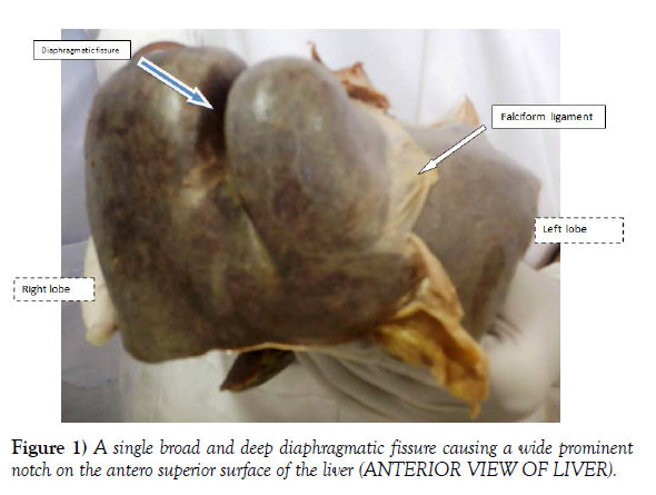
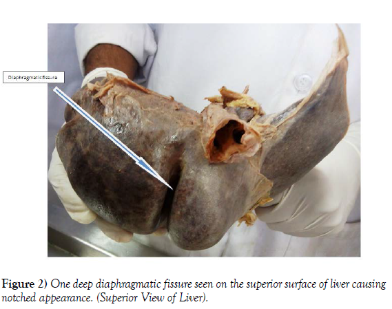
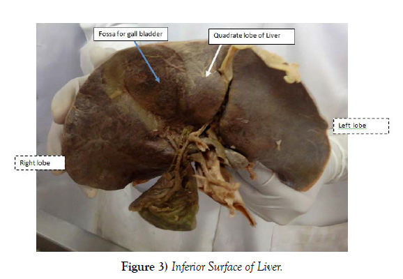
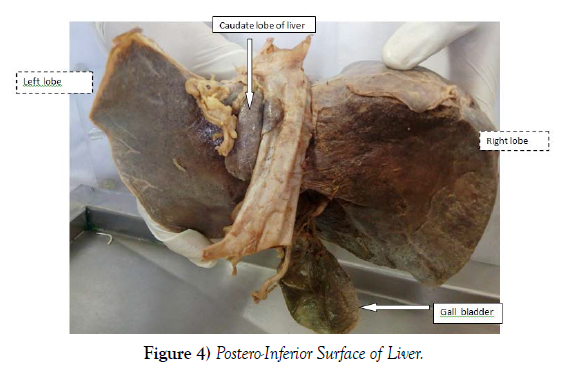
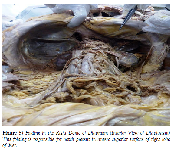
During regular dissection classes of first year medical graduates at AIIMS Bhopal, variation in the right lobe of liver was noted in a male cadever aged approximately 50-55 yrs. The liver was large and occupied whole of the right hypochondrium, upper part of the epigastrium and part of the left hypochondrium. The right lobe of the liver has one deep notch on the anterosuperior surface (Figures 1-4). Muscular diaphragmatic fold is also present in right dome of diaphragm that can be clearly seen from inferior view of diaphragm (Figure 5).
Discussion
Variations in the lobes of the liver are very rare. Presence of unusual lobes or fissures may lead to wrong radiographic diagnosis. Fitzgerald et al., [10] have reported the presence of an additional lobe. The preoperative imaging of the unusual lobe had led to the misdiagnosis as a lesser omental lymphadenopathy. Llorente and Dardik [11] have reported the presence of a large symptomatic accessory liver lobe in a 70-year-old female. The accessory lobes of liver may herniate into the thorax through the diaphragm and cause serious problems [12]. In an extensive study conducted by Joshi et al. on variations of the liver, notching along the inferior border of the caudate lobe was found in 18% of livers, a vertical fissure was found in 30% of livers, and prominent papillary process was found in 32% of cases [13]. Accessory fissures may be present rarely on the antero-superior surface of the liver [14,15]. The accessory fissures are the potential source of errors in diagnosis in imaging techniques [16]. Collection of any fluid in accessory fissures may be mistaken for a cyst, liver abscess or intrahepatic hematoma. Studies are lacking with regards to the overall shape and size of the liver. In the current case, the right lobe of the liver had two large notches and the left lobe was larger than the right lobe. The liver occupied both the hypochondria and epigastrium completely. The presence of the unusual notches on anterosuperior surface of the right lobe may lead to radiological misdiagnosis.
Conclusion
The routine anatomy text books don’t even mention about the diaphragmatic fissures and accessory sulci/ fissures. The only source of their information is the research reports. The knowledge of the diaphragmatic fissures and accessory sulci may be gainfully utilized by forensic specialists while performing postmortem study as they mimic injuries of abdominal trauma, Radiologists while interpreting Sonograms, C.T. and M.R.I. scans. Also, Confusion during hepatic resection and transplantation can be avoided by hepatobiliary surgeons.
Acknowledgement
None.
REFERENCES
- John ES, Lee JS, Panajiotis NS, Petros M, et al. Hepatic surgical anatomy. Surg Clin North Am. 2004; 84(2):413-435.
- Standring S, Ellis H, Healy JC. Gray's Anatomy: The Anatomical Basis of Clinical Practice. Elsevier Churchill Livingstone. 2005; 1213-25.
- Zahn FW. Note sur les plis respiratories du diaphragm et les sillons diaphragmatiques du foie. Rev. Med Suisse Romande. 1882; 2:531-535.
- Auh YH, Lim JH, Kim KW. Loculated fluid collections in hepatic fissures and recesses: CT appearance and potential pitfalls. Radiographics. 1994; 14(3):529-540.
- Thompson A. The morphological significance of certain fissures in the human liver. J Anat Physiol. 1899; 33(4):22.
- Macchi V, Porzionato A, Parenti A. Main accessory sulcus of the liver. Clin Anat. 2005; 18(1):39-45.
- Suksaweang S, Lin CM, Jiang TX, Hughes MW, et al. Morphogenesis of chicken liver: identification of localized growth zones and the role of beta-catenin/Wnt in size regulation. Dev Biol. 2001; 266(1):109-122.
- Newell RLM, Morgan JR. Grooves in the superior surface of the liver. Clin Anat. 1933; 18:39-45.
- Auh YH, Rubenstein WA, Ziirinsky K, Kneeland JB, et al. Accessory fissures of the liver: CT and sonographic appearance. AJR Am J Roentgenol. 1984; 143(3):565-72.
- Fitzgerald R, Hale M, Williams CR. Case report: Accessory lobe of the liver mimicking lesser omental lymphadenopathy. Br J Radiol. 1993; 66(789):839-841.
- Llorente J, Dardik H. Symptomatic accessory lobe of the liver associated with absence of the left lobe. Arch Surg. 1971; 102(3): 221-223.
- Feist JH, Lasser EC. Identification of uncommon liver lobulations. J Am Med Assoc. 1959; 169(16): 1859-1862.
- Joshi SD, Joshi SS, Athavale SA. Some interesting observations on the surface features of the liver and their clinical implications. Singapore Med J. 2009; 50(7): 715-719.
- Macchi V, Feltrin G, Parenti A, De Caro R, et al. Diaphragmatic sulci and portal fissures. J Anat. 2003; 202(3): 303-308.
- Macchi V, Porzionato A, Parenti A, Macchi C, et al. Main accessory sulcus of the liver. Clin Anat. 2005; 18(1): 39-45.
- Auh YH, Rubenstein WA, Zirinsky K, Kneeland JB, et al. Accessory fissures of the liver: CT and sonographic appearance. AJR Am J Roentgenol. 1984; 143(3): 565–572.
Indexed at, Google Scholar, Crossref
Indexed at, Google Scholar, Crossref
Indexed at, Google Scholar, Crossref
Indexed at, Google Scholar, Crossref
Indexed at, Google Scholar, Crossref
Indexed at, Google Scholar, Crossref
Indexed at, Google Scholar, Crossref
Indexed at, Google Scholar, Crossref
Indexed at, Google Scholar, Crossref
Indexed at, Google Scholar, Crossref
Indexed at, Google Scholar, Crossref
Indexed at, Google Scholar, Crossref


