Integral segmental bi-lateral renal arteries and unilateral renal veins in a cadaver: A new classification
2 Institute of Evolutionary Medicine, University of Zurich, Switzerland
Received: 05-Feb-2018 Accepted Date: Feb 13, 2018; Published: 26-Feb-2018, DOI: 10.37532/1308-4038.18.11.26
Citation: Kumaratilake JS, Saniotis A. Integral segmental bi-lateral renal arteries and unilateral renal veins in a cadaver: A new classification. Int J Anat Var. 2018;11(1):026-031.
This open-access article is distributed under the terms of the Creative Commons Attribution Non-Commercial License (CC BY-NC) (http://creativecommons.org/licenses/by-nc/4.0/), which permits reuse, distribution and reproduction of the article, provided that the original work is properly cited and the reuse is restricted to noncommercial purposes. For commercial reuse, contact reprints@pulsus.com
Summary
Right and left additional single renal arteries and additional two right renal veins were observed in a female cadaver. Additional arteries arose from the abdominal aorta and entered kidney at inferior borders of renal hila. Additional renal veins exited the kidney at the mid-posterior and inferior borders of the hilum, and joined mid-posterior and lateral wall of inferior vena cava respectively. Variations in the structure and arrangements of main and additional renal vessels have been reported, but classifications or nomenclatures used to describe them varied markedly among publications. Therefore, established a classification to unify the future descriptions. According to the new classification, the normal renal vessels are referred to as main and segmental renal arteries and veins. Additional renal arteries and veins are identified as integral segmental arteries and veins respectively. If there is more than one such vessel, they will be identified numerically from the superior to inferior poles.
Keywords
Accessory renal arteries; Additional renal arteries; Additional renal veins; Segmental renal arteries; Multiple renal arteries
Introduction
Each kidney normally received blood from a single artery that arose from the abdominal aorta (AA), entered the kidney via the renal hilum; while a single vein left the hilum, and drained the blood into the inferior vena cava (IVC) [1,2]. The right and left renal arteries (also referred to as main renal arteries) normally arose from the segment of AA between the upper and lower margins of the first and second lumbar vertebrae respectively, just below the superior mesenteric artery. Furthermore, they could arise from the AA by a common trunk [3,4]. The arrangement of arteries and veins of kidneys varied between the right and left kidneys of an individual and among individuals [1-3]. In some individuals, in addition to main renal arteries, other renal arteries originated from the AA or another artery (e.g. main renal artery) and entered the kidney at different points from upper to lower renal poles. These other arteries (i.e. additional renal arteries) have been described using different classifications and they were based on the arrangement of the additional renal arteries. These criteria and classifications are presented in Table 1.
| Criteria for classifications: | Classification of renal artery variations. | Publications | |
|---|---|---|---|
| 1) Additional renal arteries: | |||
| i) Source of origin of additional renal arteries: | [4] | ||
| a) Abdominal aorta or main renal artery | Accessary renal arteries | [4,5] | |
| b) Any artery other than AA or main renal artery (e.g. from inferior phrenic, suprarenal, ureteric, iliac, mesenteric arteries) | Aberrant or anomalous renal arteries | ||
| c) Abdominal aorta, main renal artery or any other artery (common iliac, testicular, ovarian, suprarenal, etc.) | Supernumerary renal arteries | [6] | |
| ii) Site of entry of the additional renal arteries into the kidney | |||
| a) Two renal arteries arising from the AA enter the kidney via the hilum, i.e. main renal artery and an additional renal artery. | Double hilar renal arteries. The additional hilar renal artery has been described as accessary or supernumerary hilar renal artery. | [7,4,6] | |
| b) A renal artery originating from the AA or main renal artery enters the kidney at the superior or inferior pole. | Superior or inferior polar/perforating artery. This has been described as accessary or supernumerary, superior or inferior polar/perforating artery. | [7,4,6] | |
| 2) Main renal arteries: | |||
| i) The site of origin of the right main renal artery from the AA compared to that of the left main renal artery. | The site of origin of the right main renal artery was superior to, at the same level with or inferior to that of left main renal artery | [3,5,7] | |
| ii) Branching patterns of main renal artery | Three types of arteries have been identified. | [8] | |
| a) The main renal artery – from ostium to main branching point | |||
| b) The pre-segmental artery – branch of main renal artery, which divided into two or more segmental arteries | |||
| c) Segmental arteries – branches that entered renal parenchyma. | |||
| According to the number of pre-segmental arteries, the branching patterns have been divided into 3 groups. | |||
| Type 1 – segmental arteries with no pre-segmental artery | |||
| Type 2 – segmental arteries with one pre-segmental artery | |||
| Type 3 – segmental arteries branching off from two pre-segmental arteries | |||
| Four types of variations have been described in upper segmental renal artery | [9] | ||
| iii) The course of the right main or additional renal arteries in relation to IVC. | Pre-caval renal arteries – the right main or additional renal arteries coursed anterior to the IVC. | [10] | |
AA = abdominal aorta, IVC = inferior vena cava
Table 1: Criteria and classifications that have been used for the description of renal arteries
Similar to the renal arteries, the variations in the arrangement of renal veins have been classified using following different criteria, which are presented in Table 2.
| Criteria for classifications | Classification of renal vein variations. | Publications |
|---|---|---|
| i) Site of entry of left main renal vein into the IVC compared to that of right main renal vein | The site of entry of left main renal vein was superior to, at the same level with or inferior to that of right main renal vein | [11] |
| ii) Number of renal veins | Multiple renal veins – most common renal vein variation | [5,10] |
| iii) Site of formation of main renal vein | Segmental renal veins confluence late and formed the main renal vein, i.e. more towards IVC. | [5] |
| iv) Course of the left main renal vein | a) Circumaortic left renal vein – the left main renal vein divided into anterior and posterior limbs and encircled AA. | [5] |
| b) Retroaortic left main renal vein | ||
| v) Joining of other veins into main renal veins. | a) Right gonadal and adrenal veins drained into the right main renal vein. | [5] |
| b) Retroperitoneal veins (e.g. lumbar, ascending lumbar, hemiazygos veins) drained into the left main renal vein. | [5] |
AA = abdominal aorta, IVC = inferior vena cava
Table 2: Classifications and criteria that have been used for description of additional renal veins
In early foetus, the developing mesonephros, metanephros, suprarenal glands and gonads are supplied by 9 pairs of lateral mesonephric arteries arising from the dorsal aorta. The middle group of these arteries i.e. pairs [3-5] forms renal arteries. Persistence of more than one artery pair from this group, results in additional renal arteries (as described by Budhiraja et al. [6], the findings of Felix 1912).
Knowledge on variation in the arrangement of renal arteries and veins are important, particularly in renal surgery, renal transplanting and uroradiological procedures. Therefore, the establishment of a unified method for the classification of renal artery and vein variations will be beneficial for anatomical descriptions and clinical recordings in the future.
This study, presents bilateral renal artery and unilateral renal vein variations in a cadaver, with the aim of describing a unified classification for anatomical variations in renal arteries and veins, and understands the anatomical bases for their development.
Materials and Methods
The cadaver of this study was from an eighty-three years old female, who had given her consent to donate her body to Adelaide Medical School, The University of Adelaide to be used for research and teaching. Such bodies donated to the Medical School, could be used for research by obtaining written permission from the governing committee of the body donation programme. This body was dissected to be used as a prosection for student teaching and as the body contained the variations of the renal vessels, permission was obtained from the governing committee to use the body for research. After collecting the information for research, the body has been used as a prosection for student learning. The prosection is stored in a sealed stainless-steel cabinet at 16-180C and sprayed with a wetting solution twice a week to minimize drying due to evaporation.
The body was embalmed by intra-atrial perfusion with Genelyn Anatomical mix (Adelaide, Australia) via the right common carotid artery for 3 days and stored in a 40C cold room with ultra-violet lights for six months. Skin on right and left anterior thoracic wall was reflected to about posterior axillary lines on either side after making a mid-line incision from jugular notch to xiphoid process. Anterior thoracic wall was removed by dissecting along the costal margins and xiphoid process from right to left posterior axillary lines (i.e. superior to diaphragm), cutting the ribs along the right and left posterior axillary lines and cutting the clavicles. Skin on the anterior abdomen was reflected to either side after making an incision along linea Alba from xiphoid process to pubic symphysis and extending the cuts laterally just inferior to diaphragm, and along pubic crest and inguinal ligament. Muscular wall of the anterior abdomen was also reflected to either side similarly. Intestines were dissected out from the rectum upwards, particularly, cutting the mesentery close to the intestine, in order to save the blood vessels supplying the gut. Thereafter, the peritoneum was removed to expose the kidneys, blood vessels and nerves. Then, adipose tissue and fascia were removed to identify the structures clearly and separately from one another.
Diameters and the length measurements of arteries and veins were made using a Vernier calliper. The same measurements were taken at two separate times at the same sites, by the same person. The difference between the measurements taken at the two times was calculated and the differences for each measurement were less than 3%. Thus, the measurements were considered as accurate.
Results
In this dissection, anatomical variations of the left and right renal arteries and right renal veins were seen. The detailed anatomical features of these were:
Main renal arteries
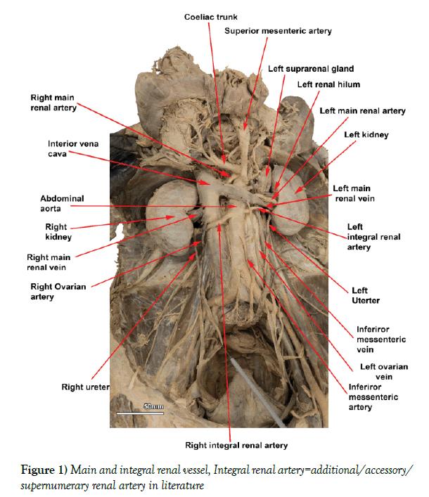
Right and left main renal arteries originated from the opposite lateral walls of the AA and the origin of the right main renal artery was 18 mm superior to that of the left main renal artery (i.e. between superior margins of right and left main renal arteries at their origin from AA) (Figure 1). The diameters of right and left main renal arteries at their origins from AA were 5.2 mm and 5 mm respectively.
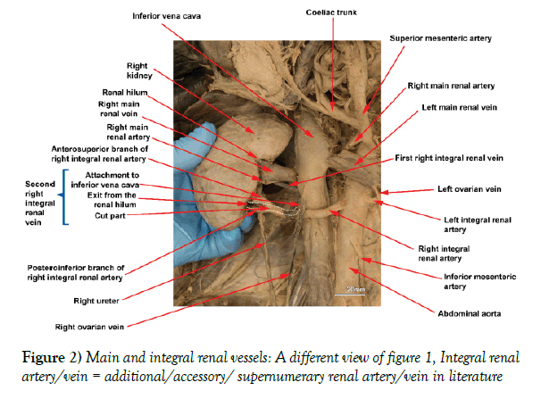
Right additional renal artery: The right additional renal artery (RARA) commenced from the right anterolateral aspect of the AA. It coursed inferior to the right main renal artery, anterior to both IVC and the right ureter, posterior to the right ovarian vein and entered the renal hilum at the inferior border (Figures 1 and 2). Just before entering the hilum, RARA divided into an anterosuperior and a posteroinferior branches (Figure 2).
Diameter of RARA at its origin from the AA was 6.5 mm. The site of origin of RARA from the AA was slightly lower than that of left additional renal artery (LARA) (i.e. 2 mm). In addition, RARA ran angled obliquely, while LARA ran more horizontally (Figure 1). The distance from the superior margin of origin of RARA from the AA to the superior margin of origin of right main renal artery was 47.2 mm. The distance from the superior margin of origin of RARA from AA to entry of anterosuperior and posteroinferior branches into the kidney at the hilum was 62 and 67 mm respectively.
Left additional renal Artery
The LARA commenced on the lateral aspect of the AA and ran inferior to the left main renal artery. This additional artery coursed posterior to the left ovarian vein and anterior to the inferior mesenteric vein and the left renal pelvis before entering the renal hilum at its inferior border. Diameter of the LARA at its origin from the AA was 3.5 mm. The distance from the superior margin of LARA at its origin from the AA to the superior margin of the origin of left main renal artery and the inferior border of left renal hilum were 22 mm and 55 mm respectively (Figure 1).
Main renal veins
Right main renal vein – shorter than the left, entered the inferior vena posterolaterally and at this location, the diameter was 10 mm. This vein ran anterior to the right main renal artery from the hilum to the inferior vena cava (Figure 1).
Left main renal vein – entered the inferior vena cava anterolaterally, superior to the right renal vein and at this site, the diameter was 14.4 mm. This vein ran posteroinferior to the left main renal artery from the hilum, close to the AA, and then coursed anterior to left main renal artery, AA and the right main renal artery before entering the IVC. The left ovarian vein joined the left renal vein anteriorly, near the inferior margin in line with the left border of AA (Figure 1).
Right additional renal veins
Two additional renal veins exited the right renal hilum and joined the IVC. The details of the veins are:
i) The first additional renal vein exited the mid-posterior margin of the right renal hilum, ran horizontally posterior to the main renal artery first and then posterior to the main renal vein and joined the mid-posterior surface of the IVC (Figure 2). The length of the vein from the mid-posterior margin of the renal hilum to the point of entry into the IVC was 51 mm and the diameter at the point of entry into the IVC was 3.2 mm.
ii) The second additional renal vein exited the right renal hilum between the two branches of RARA and joined the lateral wall of the IVC just above the joining of the right ovarian vein with the inferior vena cava (Figure 2). The vein could not be photographed as it was dissected out to demonstrate the additional renal artery and its branches. The length from the superior margin of attachment to IVC to the point of exit from the renal hilum was 35 mm. The diameter of the vein at its junction with the IVC was 6.2 mm. The distance from the superior margin of the main renal vein at its junction with the IVC to the superior margin of the additional renal vein at its junction with the IVC was 19 mm.
Discussion
The origin of right main renal artery superior to that of left main renal artery (Figure 1) has been described [3,5,7]. The incidences of origin of right main renal artery superior to, at the same level with and inferior to that of left main renal artery from the AA have ranged from 53.8–37%, 50–34.6% and 13.0–11.5% individuals respectively [3,7]. Therefore, the origin of right main renal artery either superior to as in the current case (Figure 1) or at the same level with that of left main renal artery from the AA, has ranged from a common anatomical variation to a normal anatomical arrangement.
The RARA and LARA in the current study, originated from the AA and entered the kidneys via the hila together with the main renal arteries, thus they represent the double hilar artery category previously described [5-9]. In addition, the RARA ran anterior to the IVC, thus it also belongs to the pre-caval group as described by Bouliet et al., (Figures 1 and 2) [10].
The anatomy and the arrangement of the right and left main renal veins appeared to be normal. However, the passage of the left main renal vein posteroinferior to the left main renal artery from the hilum to close to the left border of AA may represent a minor variation (Figure 1); because the renal veins usually lied anterior to the renal artery at the renal hilum [5]. The two additional renal veins seen in association with the right kidney (Figure 2) represent the most common renal venous variation of the right side of the body and was evident in approximately 15-30% of the population [5].
Additional renal arteries have been described as end arteries [4,6], similar to the segmental arteries of the kidney [9], thus the former arteries are the sole arterial supply to the respective segments of the kidney (i.e. not accessory, additional or supernumerary). In other words, additional renal arteries are an integral part of the arterial supply of the kidney and represent the segmental arteries of those segments. During foetal development, these were the segmental arteries of the foetal kidney at a particular stage of development, but did not get removed as the development progressed.
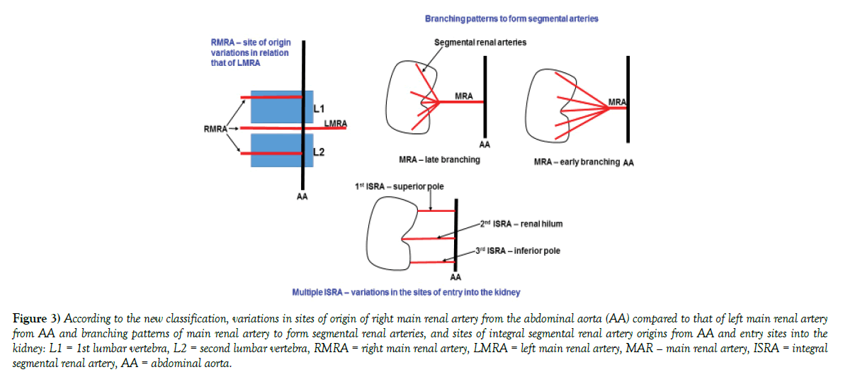
Furthermore, new segmental arteries would not have grown into these segments from the main renal artery as foetal development progressed. Therefore, the additional renal arteries remained as the adult functional segmental arteries of those renal segments. Therefore, the use of terminologies such as accessary, additional, aberrant [4] or supernumerary renal arteries [6], and first and second additional renal arteries [11] to identify the variations of the renal arteries are not appropriate. Therefore, the following classification has been proposed to identify and describe renal arteries, to unify the description of renal arteries. This classification is presented in Table 3 and Figure 3.
| Renal vessel | New Classification |
|---|---|
| Consider percentage of incidence of 50% or more as normal and less than 50% as variation. | |
| Arteries: | |
| i) Right or left main renal artery, which is some-times referred to as right or left normal renal artery [5] | Continue to refer as the right or left main renal artery, as it supplies the full kidney or a major part of the kidney, when additional renal arteries are present. |
| Variations – describe: | |
| i) According to the origin of: | |
| a) The right main renal artery in relation to that of the left main renal artery from the AA (i.e. superior to, at the same level with or inferior to). | |
| b) Right or/and left main renal artery/arteries arise from the AA by a common trunk or from an artery other than AA. | |
| ii) Branching pattern to form segmental arteries; | |
| a) Early branching of main renal artery – i.e. closer to AA. | |
| b) Late branching of main renal artery – i.e. closer to hilum or at hilum. | |
| c) Branching via pre-segmental arteries to form segmental arteries as described by Kang et al., [8]. | |
| iii) The course of the right main renal arteries in relation to IVC – i.e. pre-caval. | |
| ii) Segmental renal arteries | Continue to identify the normal segmental renal arteries as segmental renal arteries. as they continue to solely supply a renal segment |
| Variations: | |
| Additional segmental renal arteries should be referred to as integral segmental renal arteries, as they continue to solely supply a renal segment. Describe them: | |
| i) According to the artery of origin i.e. origin from AA, main renal artery or any other artery (inferior phrenic, suprarenal, ureteric, iliac, mesenteric, testicular, ovarian) | |
| ii) According to the site of entry into the kidney (i.e. superior pole, hilum or inferior pole) | |
| If there are more than one integral segmental renal artery per kidney, identify them numerically from the superior to inferior poles of the kidney |
AA = abdominal aorta, IVC = inferior vena cava
Table 3 The identification of adult renal arteries (i.e. normal and additional) according to the new proposed classification
Figure 3) According to the new classification, variations in sites of origin of right main renal artery from the abdominal aorta (AA) compared to that of left main renal artery from AA and branching patterns of main renal artery to form segmental renal arteries, and sites of integral segmental renal artery origins from AA and entry sites into the kidney: L1 = 1st lumbar vertebra, L2 = second lumbar vertebra, RMRA = right main renal artery, LMRA = left main renal artery, MAR – main renal artery, ISRA = integral segmental renal artery, AA = abdominal aorta.
According to this classification, the integral segmental renal arteries in the current study could be described as:
i) Right integral segmental renal artery originated from the AA, inferior to the right main renal artery and entered the right kidney at the lower border of hilum. Before entering into the hilum, the artery divided into an anterosuperior and a posteroinferior branches (Figure 2) and the length of the artery from the AA to hilar end of the two branches was 62 mm and 67 mm respectively. Diameter of the artery at its origin was 6.5 mm
ii) Left integral segmental renal artery originated from the AA, inferior to left main renal artery and entered the left kidney at the lower border of the hilum (Figure 1). Length of the artery from the AA to the hilar end was 55 mm. Diameter of the artery at its origin was 3.5 mm.
The measurement of the length (i.e. from origin to site of entry into the kidney) and the diameter (i.e. at site of origin) of the segmental renal arteries including the integral segmental renal arteries will be of value in surgical settings, where clamping of the segmental arteries will be required [8].
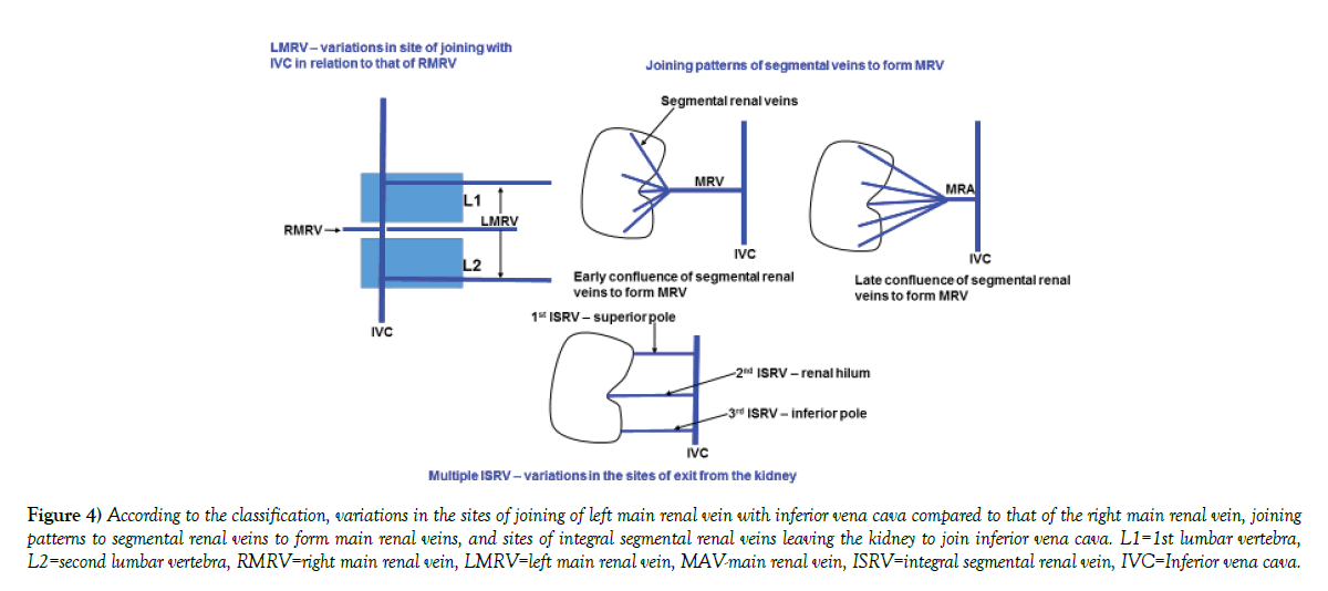
Like the segmental renal arteries, the segmental renal veins primarily drain the respective renal segments, even though there is some collateral venous circulation between adjacent renal segments [12]. Therefore, an additional renal vein is also an integral part of the venous circulation of the kidney (i.e. each draining a renal segment), thus could be considered as an integral segmental renal vein. Therefore, the following classification has been proposed to identify and describe renal veins, to unify the description of renal veins. This classification is presented in Table 4 and Figure 4.
| Renal vessel | New Classification |
|---|---|
| Consider percentage of incidence of 50% or more as normal and less than 50% as variation. | |
| Veins: | |
| i) Right or left main renal vein, which is some-times referred to as right or left normal renal vein | Continue to refer as the right or left main renal vein, as it drains the full kidney or a major part of the kidney, when additional renal veins are present. |
| Variations – describe: | |
| i) According to the joining of: | |
| a) The left main renal vein in relation to that of the right main renal vein with IVC (i.e. superior to, at the same level with or inferior to). | |
| b) Right or/and left main renal vein/veins may join to froma common trunk that joins with IVC or a vein other than IVC.. | |
| ii) Joining pattern of segmental veins to form the main renal vein. | |
| a) Early joining of segmental renal veins – i.e. long main renal vein. | |
| b) Late joining of segmental renal veins– i.e. short main renal vein | |
| iii) The course of the left main renal vein in relation to AA – i.e. Circumaortic left main renal vein; retroaortic left main renal vein | |
| iv) Joining of other veins into main renal veins. – i.e. a) Right gonadal and adrenal veins draining into the right main renal vein. b) Retroperitoneal veins (e.g. lumbar, ascending lumbar, hemiazygos veins) draining into the left main renal vein. | |
| ii)Segmental renal veins | Continue to identify the normal segmental renal veins as segmental renal veins as they continue to “solely” drain a renal segment |
| Variations: | |
| Additional segmental renal veins should be referred to as integral segmental renal veins, as they continue to solely drain a renal segment. Describe them: | |
| i) According to the site of confluence to form the main renal vein. That is early near the hilum and late near the IVC. | |
| ii) According to the site of exit from the kidney (i.e. superior pole, hilum or inferior pole) | |
| iii) Number of integral segmental renal vein per kidney. Identify them numerically from the superior to inferior poles of the kidney |
IVC = inferior vena cava, AA = abdominal aorta
Table 4: Integral segmental renal vein classification
Figure 4) According to the classification, variations in the sites of joining of left main renal vein with inferior vena cava compared to that of the right main renal vein, joining patterns to segmental renal veins to form main renal veins, and sites of integral segmental renal veins leaving the kidney to join inferior vena cava. L1=1st lumbar vertebra, L2=second lumbar vertebra, RMRV=right main renal vein, LMRV=left main renal vein, MAV-main renal vein, ISRV=integral segmental renal vein, IVC=Inferior vena cava.
According to this classification, in the current case, the integral segmental renal veins in the right kidney could be described as:
i) First right integral segmental renal vein, which exited the right kidney at the middle of the posterior border of the renal hilum, ran horizontally, first posterior to main renal vein and the artery, then posterior to the main renal vein and joined the IVC on its mid-posterior surface. This vein may represent the tributary draining the posterior renal segment. The length and the diameter of the vein were 51 mm and 3.2 mm respectively.
ii) Second right integral segmental renal vein, which exited the right kidney at the inferior border of the hilum, between the two branches of the right integral segmental renal artery, ran at an angle superiorly and joined IVC anterolaterally, just superior to the joining of right ovarian vein (Figure 2). The length and the diameter of the vein were 35 mm and 6.2 mm respectively.
Like the integral segmental renal arteries, the length (i.e. from the site exiting the kidney to the point of joining with the draining vein) and the diameter (i.e. at the point of joining with the draining vein) of the integral segmental renal veins may be of value in surgical situations.
Above simple classifications could bring uniformity into anatomical descriptions and clinical reporting of the integral segmental renal arteries and veins.
Conclusion
Variations in the arrangement of renal vessels have been reported and are a very important aspect in many clinical intervention of the kidney, such as renal transplantation, repair of aneurisms of abdominal aorta, and urological and angiographic interventions. In literature dealing with anatomical variations of the vessels of the kidney, the classifications and the terminology used to describe variations differ substantially among publications, thus there is no uniformity in published information. Current publication presents a new classification based on the anatomy of the renal blood supply and it could be applied to most of the anatomical variations reported in relation to renal vessels. Therefore, this classification may help to introduce some uniformity into the reporting and recording of anatomical variations of renal vessels thus may be useful to clinicians and anatomists in the future. The concept introduced here that additional renal arteries are end arteries, thus not accessary, supernumerary, aberrant or additional arteries, but integral part of the renal circulation. This indicates that, the variant arteries are supplying specific renal segments, thus they are segmental arteries of different embryonic origin.
REFERENCES
- Reginelli A, Izzo A, D’Andrea A, et al. Renovascular anatomic variants at CT angiography. Int Angiol. 2015;34:36-42.
- Hassan SS, Johnson JC, Ettarh R, et al. Incidence of variations in human cadaveric renal vessels. Folia Morphol. 2017;76:394-407.
- Beregi JP, Willoteaux S, Remy-Jardin M, et al. Anatomic variation in the origin of the main renal arteries: spiral CTA evaluation. Eur Radiol. 1999;9:1330-34.
- Saritha S, Jyothi N, Supriya G, et al. Cadaveric study of accessory renal arteries and its surgical correlation. Int J Res Med Sci. 2013;1:19-22.
- Turkvatan A, Cumhur T, Olcer T, et al. Multidetector CT angiography of renal vasculature: normal anatomy and variants. Eur Radiol. 2009;19:236-44.
- Budhiraja V, Anjankar V, Goel P, et al. Supernumerary Renal Arteries and Their Embryological and Clinical Correlation: A Cadaveric Study from North India. ISRN Anatomy.
- Cicekcibasi AE, Salbacak A, Buyukmumcu M, et al. An investigation of the origin, location and variations of the renal arteries in human foetuses and their clinical relevance. Ann Anat. 2005;187:421-27.
- Kang WY, Park BJ, Han NY, et al. Perihilar branching patterns of renal artery and extrarenal length of arterial branches and tumour-feeding arteries on multi detector CT angiography. Br J Radiol. 2013.
- Mishra GP, Bhatnagra S, Singh B. Anatomical variations of upper segmental renal artery and clinical significance. J Clin Diagn Res. 2015;9:AC01-3.
- Bouali O, Molinier F, Benouaich V, et al. Anatomic variations of the renal vessels: focus on the precaval right renal artery. Surg Radiol Anat. 2012;34:441-6.
- Satyapal KS. Intra renal angles, entry into inferior vena cava and vertebral levels of renal veins. Anat Rec. 1999;258:202-7.
- Sinnatamby CS. Last’s anatomy, Regional and applied. London, New York, Sydney: Churchill; Livingstone, Elsevier. 2011;284.


