Squatting facets of talus in the coastal population of Mangalore
2 Department of Anatomy, SRM Medical College Hospital and Research Centre, Bangalore, India
3 Department of Anatomy, Kasturba Medical College, Mangalore, India
Received: 12-Dec-2019 Accepted Date: Jan 06, 2020; Published: 13-Jan-2020, DOI: 10.37532/1308-4038.20.13.73
Citation: Bhat K, Balakrishnan R, Saralaya VV. Squatting facets of talus in the coastal population of mangalore. Int J Anat Var. 2020;13(1): 73-75.
This open-access article is distributed under the terms of the Creative Commons Attribution Non-Commercial License (CC BY-NC) (http://creativecommons.org/licenses/by-nc/4.0/), which permits reuse, distribution and reproduction of the article, provided that the original work is properly cited and the reuse is restricted to noncommercial purposes. For commercial reuse, contact reprints@pulsus.com
Abstract
The development of bipedal locomotion is one of the most significant adaptations of the hominin lineage and the foot is particularly specialized for this purpose. Talus is one of the most important members of the tarsal bones as it carries the weight in stationary position and during movements. The articular morphology of the human skeleton can be subject to modification by stresses imposed upon it and habitual squatting alters the skeletal morphology of the lower limb.
Aim: To study the frequency of squatting facets and trochlear extensions in tali and their regional peculiarity.
Methods: This is a descriptive study done on 96 human’s dry tali of unknown age and sex obtained from the Department of Anatomy. Each talus was examined in detail for the presence of squatting facets and trochlear extensions.
Results: Squatting facets and trochlear extensions were observed in (55, 57.3%) tali and were more common on right tali (33, 70.2%). Majority of the tali were observed to have medial facet (33, 34.4%) and medial extension (28, 29.2%) alone and a combination of medial facet and medial extension was observed in (20, 20.8%) tali.
Conclusion: Medial facet and medial extension both alone and in combination were frequently observed and were common on the right tali than left tali. Squatting facets and trochlear extensions were common in our study on Indians than the Europeans due to the habitual squatting position.
Keywords
Human skeleton; Orthopedic surgeons; Anthropologists
Introduction
The foot is particularly specialized both anatomically and functionally for bipedal locomotion and subsequently undergoes strong selection pressure to deal with both balance and propulsion in a highly efficient way [1].
In stationary position and during movement Talus is one of the important bone as it carries the weight of the entire body. Its main importance lies in the fact that it forms a connecting link between the bones of leg and foot and undergoes a lot of stress due to routine day to day activity. Its integrity is vital for all locomotor movements. It is also unique amongst all the bones in the foot by virtue of its total absence of any muscular attachments and tenuous blood supply [2].
As all bones a Talus bears the head, neck, and body head articulates with the navicular bone, the body articulates with distal end of tibia 2, neck makes an angle of 150 degrees with the body and ‘Squatting facet’ are commonly present on the dorsolateral surface of the neck of Talus.
Squatting is a resting postural complex involving hyperextension at the hip and knee, dorsiflexion at the ankle and sub-talar joint [3]. Skeletal morphology of the lower limb has been recognised to be subjected to modifications by the stress upon it.
The anterior margin of the lower end of the Tibia is bevelled due to extreme dorsiflexion of the ankle, the lower end of the Tibia articulates with the facet or facets on the dorsal aspect of the neck of the talus. They may be lateral or medial in position and usually the lateral one is often continuous with the trochlear articular surface [4].
Modifications at the neck of the Talus and the distal Tibia are subjected to mechanical stress induced by the squatting habit observed in our population. In spite of being such an important bone to the anatomists, orthopaedic surgeons, anthropologists, and forensic experts, there is a dearth of literature available on its morphological features in Indians which needs to be assessed.
Aims and Objectives
➣ To study the frequency of trochlear extension and squatting facets in Tali.
➣ To study the regional peculiarity of trochlear extension and squatting facet.
Materials and Methods
This was a descriptive study done on 96 human dry tali of unknown age and sex from the coastal population of Mangalore, obtained from the Department of Anatomy, Kasturba Medical College, Centre of Basic Sciences, Bejai, Mangalore from April 2013 to June 2013, after obtaining Institute Ethics Committee clearance.
Tali which were free of pathological changes or anomalies were taken for the study. The tali were first identified as right and left and were numbered and photographed. The presence of squatting facets and trochlear extensions in the Tali were examined in detail.
Statistical analysis
The data collected were tabulated in Microsoft Excel Worksheet and computer-based analysis was performed using the SPSS 16.0 software (SPSS, Chicago, IL, USA).
Results
Ninety-six human dry tali were studied, of which (47, 49%) were right tali and (49, 51%) were left tali.
Squatting facets and trochlear extensions
Squatting facets and trochlear extensions were observed in (55, 57.3%) tali and were more common on right tali (33, 70.2%). No squatting facets and trochlear extensions were observed in (41, 42.7%) tali. The majority of the tali were observed to have medial facet (33, 34.4%) and medial extension (28, 29.2%). The frequency distribution of squatting facets and trochlear extensions are tabulated in (Table 1). Distribution of squatting facets and trochlear extensions based on the side of tali are shown in (Table 2).
TABLE 1 Distribution of squatting facets and trochlear extensions.
| Parameter | No. of tali n (%) |
|---|---|
| Medial facet | 33 (34.4) |
| Lateral facet | 29 (30.2) |
| Medial extension | 28 (29.2) |
| Lateral extension | 13 (13.5) |
TABLE 2 Distribution of squatting facets and trochlear extensions based on side of tali.
| Parameter | No. of Right tali n (%) | No. of Left tali n (%) |
|---|---|---|
| Medial facet | 18 (38.3) | 15 (30.6) |
| Lateral facet | 19 (40.4) | 10 (20.4) |
| Medial extension | 18 (38.3) | 10 (20.4) |
| Lateral extension | 9 (19.1) | 4 (8.2) |
Combination of squatting facets and trochlear extensions Most of the tali (20, 20.8%) had a combination of medial facet and medial extension and were common on the right tali. The distribution of the combination of squatting facets and trochlear extensions and their distribution based on the side of tali are shown in (Tables 3 & 4) respectively.
TABLE 3 Distribution of combination of squatting facets and trochlear extensions.
| Parameter | No. of tali n (%) |
|---|---|
| Medial facet and medial extension | 20 (20.8) |
| Medial facet and lateral facet | 12 (12.5) |
| Lateral facet and lateral extension | 11 (11.5) |
| Medial extension and lateral extension | 9 (9.4) |
TABLE 4 Distribution of combination of squatting facets and trochlear extensions.
| Parameter | No. of Right tali n (%) | No. of Left tali n (%) |
|---|---|---|
| Medial facet and medial extension | 11 (23.4) | 9 (18.4) |
| Medial facet and lateral facet | 8 (17) | 4 (8.2) |
| Lateral facet and lateral extension | 7 (14.9) | 4 (8.2) |
| Medial extension and lateral extension | 6 (12.8) | 3 (6.1) |
Discussion
Talus is the second largest of the tarsal bones takes part in the formation of talocrural, subtalar and talocalcaneonavicular joints [5,6]. Squatting facets on the dorsal surfaces of the neck of the Talus in the foetus of oriental races provide evidence for inheriting acquired characters. Barnett also suggested that Squatting facets are more commonly seen in European foetuses than in adults [6]. Evidences suggest that Indian foetus inherit no greater expression of squatting facets than the European fetus and the fetal presence of such facets appears likely to result from the considerable dorsiflexion of the foot that occurs during intrauterine life [3].
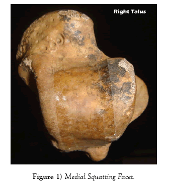
‘Medial squatting facet’ does not follow the line of curvature of the trochlear surface and is separated from the surface by a transverse ridge of bone, medial squatting facet is present on the upper medial surface of the neck (Figure 1). This facet does not articulate with the tibia in dorsiflexion. It appears to be associated with the habit of squatting [6].
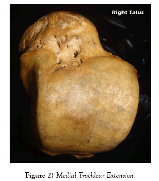
‘Medial extension of the trochlear surface’ (Figure 2). It is invariably associated with a forward prolongation of the medial articular surface of the talus [6].
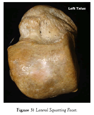
‘Lateral squatting facet’ present on the lateral surface of the neck of the talus, articulating in full dorsiflexion with the anterior surface of the lower border of tibia (Figure 3). It is often divided from the anterior margin of the trochlea by a distinct groove; in other tali, it is continuous with the trochlear surface, though makes a sharp angle with the line of curvature of the latter [6].
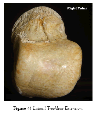
Anterior margin of the trochlear surface of the talus exhibits a sinuous form due to forward extension of the lateral one-third of this surface. This extension continues the line of curvature of the trochlea and thus makes contact with the under surface of the tibia during dorsiflexion. Lateral extension of the trochlear surface is not a true squatting facet and hence the term is coined so (Figure 4) [6].
The adequate knowledge of the anatomical variations of the talus is significant, especially in Indians, due to the habitual squatting position as it can be of help for foot rehabilitation and reconstruction.
In our study, we observed trochlear extension and squatting facets to be present in (55, 57.3%) tali and were commonly identified on the right tali (33, 70.2%). No squatting facets and trochlear extensions were seen in (41, 42.7%) tali which were less when compared to that reported by Oygucu et al. (65, 37.1%) [3].
Individually, medial facet and medial extension were commonly observed in our study. Conversely, Oygucu et al. [3] in a study on trochlear extension and squatting facets on the neck of talus in late byzantine males observed lateral squatting facets to be the commonest who was also frequently observed in studies done by Das [7] and Singh [8]. These studies [3,7,8] reported medial extension to be common than lateral extension similar to the present conducted study.
The prevalence of medial squatting facet (33, 34.4%) in the present study was greater than that reported by Oygucu et al. (1, 0.6%), and Das (8, 4%) but less than that observed by Pandey and Singh (46, 17.6%) [3,7,9]. We had a higher prevalence of medial extension (28, 29.2%) than that observed by Barnet [6] in his study on Europeans (11, 11%) and less than that reported by Singh [8] in his study on Indians (74, 24.6%).
In our study, the occurrence of lateral squatting facet (29, 30.2%) was greater than that reported by a European study (2, 2%) but was less when compared to other Indian studies [6-8]. The prevalence of lateral extension (13, 13.5%) observed in our study was less when compared to other studies [6-9].
We observed a combination of medial facet and medial extension (20, 20.8%) to be more frequent than other combinations and all the combinations were commonly seen on the right tali. Medial and lateral facet together was observed in (12, 12.5%) tali which were greater than that reported by Oygucu et al. (1, 0.6%) and Das (3%) followed by lateral extension and lateral facet (11, 11.5%) and medial and lateral extension together (9, 9.4%) [3,7].
It has been observed that the prevalence of medial extension and squatting facets in the study were higher than that of the late Byzantine [3] and Europeans [6] but was less than that reported in other Indians studies [7-9]. The prevalence of lateral extension in our study was less when compared to other studies [6-9].
Similar to the present study, the study by Singh on squatting facets on talus and tibia on Indian population, he observed that squatting facets were more common in the Indian adult as compared to the European adult and Indian fetus and in the European fetus as compared to the European adult and Indians [8].
The reason for the variations in the present study and others may be due to the fact that it is unlikely, that precisely the same factors determine the expression of trochlear extension and squatting facet. In Indians, the squatting facets and trochlear extensions are common than other races and they persist into adult life due to the habitual squatting position the Indians maintained throughout their lifetime.
Conclusion
In our study medial facet and medial extension, both alone and in combination were frequently observed and were common on the right tali than left tali. Squatting facets and trochlear extensions were common in our study on Indians than the Europeans due to the habitual squatting position. Unlike the weight-bearing articular facets of the major weight bearing bone of the body such as femur, pelvis and tibia the talar articular surfaces are very small in relation to the loading environment and their ability to remain morphologically plastic during ontogeny represents an adaptation, that is integral to our ability to cope with the substantial gains in body weight that occur during our developmental period. Adequate knowledge of the occurrence and variations in the facets and extensions in tali may be useful for the reconstructive procedures carried out in the foot.
REFERENCES
- Smith WEHH, Aiello LC. Fossils, feet and the evolution of human bipedal locomotion. J Anat. 2004;204:403-16.
- Standring S. Gray’s anatomy- The anatomical basis of clinical practice, 40th edn., Spain: Elsevier Limited. 2008:1429-62.
- Oygucu H, Kurt MA, Ikiz I, et al. Squatting facets on the neck of the talus and extensions of the trochlear surface of the talus in late byzantine males. J Anat. 1998;192:287-91.
- Frazer EJ. Lower extremity: Tibia, fibula and foot. In: Breathnach AS, eds. Frazer’s Anatomy of the human skeleton. 6th edn., Great Britain: J & A Churchill limited. 1965:132-61.
- Kaur M, Kalsey G, Laxmi V. Morphological classification of tali on the basis of calcanean articular facets. Pb J Orthop. 2011;12:57-60.
- Barnett CH. Squatting facets on the European talus. J Anat. 1954;88:509-13.
- Das AC. Squatting facets of the talus in U.P. subjects. J Anat Soc India. 1959;8:90-2.
- Singh I. Squatting facets on the talus and tibia in Indians. J Anat. 1959;93:540-50.
- Pandey SK, Singh S. Study of squatting facet/extension of talus in both sexes. Med Sci Law. 1990;30:159-64.


