Unusual origin of the left vertebral artery
Nurcan Imre*, Bulent Yalcin and Hasan Ozan
Department of Anatomy, Gulhane Military Medical Academy, Ankara, Turkey
- *Corresponding Author:
- Nurcan Imre, MD
Assistant Professor, Department of Anatomy, Gulhane Military Medical Academy (GATA), 06018 Etlik-Ankara, Turkey
Tel: +90 (312) 304 3503
E-mail: nercikti@gata.edu.tr
Date of Received: October 20th, 2009
Date of Accepted: February 2nd, 2010
Published Online: May 17th, 2010
© Int J Anat Var (IJAV). 2010; 3: 80–82.
[ft_below_content] =>Keywords
vertebral artery, variations, aortic arch, subclavian artery
Introduction
The vertebral artery (VA) is important to posterior cerebral circulation. It arises from the superior surface of the first part of the subclavian artery medial to the scalenus anterior muscle. The vessel takes a vertical posterior course to enter into the transverse process of the sixth cervical vertebra. It continues through the transverse foramina of the cervical vertebrae and after passing through the transverse foramen of the atlas, turns posteromedially on its posterior arch, pierces the atlantooccipital membrane and the dura mater, respectively and then enters the foramen magnum [1,2]. The segment of the VA from its origin at the subclavian artery to its entry into the respective transverse foramina is called the pretransverse or prevertebral segment [2]. Several researchers have reported anomalous origins of the VA such as from the aortic arch, between the left common carotid artery (LCCA) and left subclavian artery (LSA) or after LSA, from the thyrocervical trunk, from the brachiocephalic trunk (BT), from the common carotid artery, from the external carotid artery, from a common carotid trunk formed by LSA and left vertebral artery (LVA) [3–7]. Knowing the variation of the origin of VA and its prevertebral course is of great importance for head and neck surgery [8] and angiography [2] and vascular angiography.
Case Report
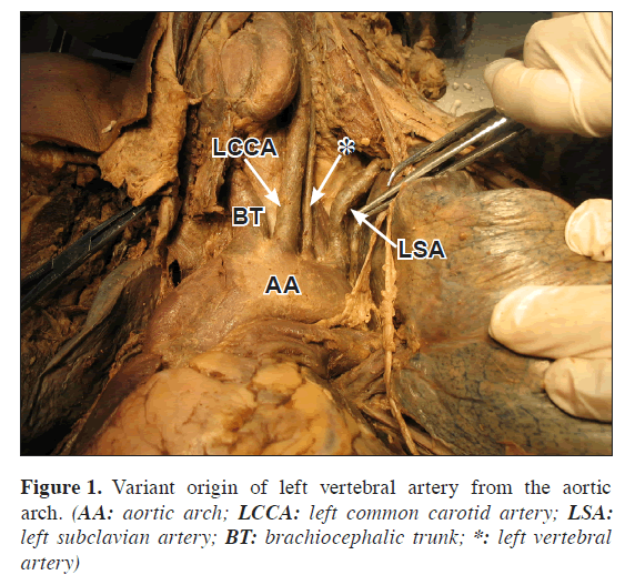
During a routine dissection of the branches of the aortic arch of a 81-year-old female cadaver at the Faculty of Medicine, Gulhane Military Medical Academy, we encountered a variation of the aortic arch gave off four branches, BT, LCCA, LVA, LSA. The LVA originated from the aortic arch between the origins of the LCCA and the LSA. The origins distance between the LVA and the arteries were 3.96 mm and 5.10 mm, respectively. Diameter of the LVA at its origin was 5.51 mm. The LVA coursed upward to the transverse foramen of the C6. The length of the prevertebral segment of the LVA was 88.5 mm. The right vertebral artery (RVA) originated from the right subclavian artery. Its origin was 6.84 mm distal from the origin of the right subclavian artery (RSA). Its diameter at its origin was 4.54 mm. The artery entered the transverse foramen of the C6. The length of the prevertebral segment of the RVA was 44.31 mm.
Discussion
The intersegmental arteries are 30 or more branches of the dorsal aorta [1]. They originate from the branchial aortic system. Initially the VA emerges being formed by the longitudinal anastomosis of the cervical segmental arteries [9,10]. Usually the first part of vertebral artery develops from proximal part of dorsal branch of seventh cervical intersegmental artery proximal to postcostal anastomosis. The second part is derived from longitudinal communications of the postcostal anastomosis. In the present case, the left sixth dorsal intersegmental artery might have persisted as the first part of vertebral artery hence LVA wass arising from arch of aorta.
Origin point of the VA were reported as from the aortic arch, between the LCCA and LSA or after LSA, from the thyrocervical trunk, from the brachiocephalic trunk, from the common carotid artery, from the external carotid artery, from a trunk formed by LSA and LVA. [3–7].
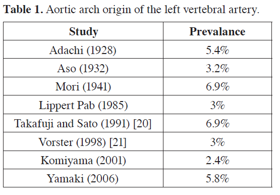
Incidence of the LVA originating from the aortic arch was reported variable (Table I). The origin of the VA and the entrance point into the cervical transverse foramen were examined during dissection of 40 cadavers at the Faculty of Medicine, Gulhane Military Medical Academy from 2000 to 2009. In one female body the LVA was originated from the aortic arch between the origins of the LCCA and the LSA. The frequency of a LVA arising from the aortic arch was found as 2.5%.
There are many reports in the literature of frequency of the aortic arch origin of the LVA (Table 1). It ranges from 2.4 % to 6.9 %. Nevertheless, most authors have stated that it is about 2.5-3 %.
Lippert Pab’s classified the LVA according the origin from the aortic arch as: between the LCCA and LSA (Type A, 3%), between a common trunk formed by BT and LCCA and LSA (Type B, <1%), after the LSA (Type C, <1%), after the LSA as the third branch (Type D, <0,1%), after a common trunk as the second branch (Type E, <0,1%), different from Type A, RSA appears from descending aorta (Type F, <0,1%), one of two roots as a penultimate branch (Type G, <1%), both VA branch from the aortic arch (Type H, <0,1%) [11].
Nizanowski, in their study on 160 cadavers and 100 fetuses, found the LVA originating from the aortic arch in seven adults and one fetus [12].
Komiyana reported the incidence of arterial dissection of the vertebral artery of aortic origin and vertebral artery of subclavian origin [13]. According to their studies LVA of aortic origin was associated with a significantly higher incidence of vertebral artery dissection than LVA of left subclavian artery origin and right vertebral artery of right subclavian origin [13].
Gluncic reported the left vertebral artery originated from the common trunk of it and the left subclavian artery at the aortic arch [3]. Its origin was 3.5 mm distal from the origin of the trunk. The length of the prevertebral segment of the left vertebral artery was 87.3 mm and its diameter at the origin was 3.3 mm [3]. Panicker reported the LVA originated directly from arch of aorta between the left common carotid artery and left subclavian artery [14]. The diameter of left vertebral artery at origin was 3.1 mm as compared to that of the right, which had a diameter of 6.5 mm at origin. The length of right vertebral artery was 38 mm and that of left vertebral artery was 92 mm [14]. In the present case, diameter of the LVA at its origin was 5.51 mm. The length of the prevertebral segment of the LVA was 88.05 mm.
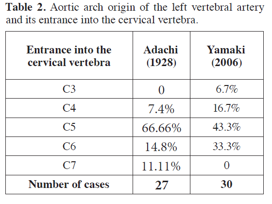
Entrance point of the LVA originating from the aortic arch was also reported variable (Table 2). Most common entrance points were reported as C5 and C6, respectively [15,16]. Nevertheless, C6 reported as the most common entrance point [16].
Overall, the most common entrance for vertebral arteries was at C6; 82.1% for left vertebral arteries [16]. Different levels of entry of the VA to the transverse foramen may also contribute to differences in hemodynamics [13].
The anatomic and morphologic variations of the LVA are significant for diagnostic and surgical procedures in the head and neck region [2,3,17]. It is of clinical importance to know the origin and course of prevertebral segment of the vertebral artery in detail and being aware of the possible variations. In order to prevent complications, it is critical to assess vascularization in this region prior to conducting medical procedures. The extracranial portion of VA is frequently affected from atherosclerosis. The most common site of the consequent stenosis is its origin. According to Bernardi and Detori, the unusual origin of the VA ‘‘may favor cerebral disorders because of alterations in the cerebral haemodynamics’’[18].
Goray et al. reported a variation in the origin of VA, where the case was asymptomatic. Some patients complained of dizziness, however, which was thought to have no connection to the anomalous origin of the VA. Anomalous VA origin also represents a potential pitfall in diagnostic cerebrovascular injury [19].
References
- Moore KL. The Developing Human. Clinically Oriented Embryology. 3rd Ed. WB Saunders, Philadelphia. 1982; 291–318.
- Matula C, Trattnig S, Tschabitscher M, Day JD, Koos WT. The course of the prevertebral segment of the vertebral artery: anatomy and clinical significance. Surg Neurol. 1997; 48: 125–131.
- Gluncic V, Ivkic G, Marin D, Percac S. Anomalous origin of both vertebral arteries. Clin Anat. 1999; 12: 281–284.
- Mahmutyazicioglu K, Sarac K, Boluk A, Kutlu R. Duplicate origin of left vertebral artery with thrombosis at the origin: color Doppler sonography and CT angiography findings. J Clin Ultrasound. 1998; 26: 323–325.
- Nonami Y, Tomosawa N, Nishida K, Nawata S. Dissecting aortic aneurysm involving an anomalous right subclavian artery and isolated left vertebral artery: Case report and review of the literature. J Cardiovasc Surg (Torino). 1998; 39: 743–746.
- Strub WM, Leach JL, Tomsick TA. Left vertebral artery origin from the thyrocervical trunk: a unique vascular variant. AJNR Am J Neuroradiol. 2006; 27: 1155–1156.
- Yazar F, Yalcin B, Ozan H. Variation of the aortic arch branches: Two main trunks originating from the aortic arch. Gazi Medical Journal, 2003; 14: 181–184.
- Krmpotic-Nemanic J, Draf W, Helms J. Surgical anatomy of head and neck. Berlin, Springer-Verlag. 1985; 30.
- Haughton VM, Rosenbaum AE. The normal and anomalous aortic arch brachiocephalic arteries. In: Newton TH, Potts DG, eds. Radiology of the skull and brain. St. Louis, Mosby. 1978; 1145–1163.
- Newton TH, Mani R. The vertebral artery. In: Newton TH, Potts DG, eds. Radiology of the skull and brain. St. Louis, Mosby. 1978; 1659–1709.
- Lippert H, Pabst R. Arterial Variations in Man. Classification and Frequency. JF Bergmann Verlag, Munchen. 1985; 30–38.
- Nizanowski C, Noczynski L, Suder E. Variability of the origin of ramifications of the subclavian artery in humans (studies on the Polish population). Folia Morphol. (Warsz). 1982; 41: 281–294.
- Komiyama M, Morikawa T, Nakajima H, Nishikawa M, Yasui T. High incidence of arterial dissection associated with left vertebral artery of aortic origin. Neurol Med Chir (Tokyo). 2001; 41: 8–11.
- Panicker HK, Tarnekar A, Dhawane V, Ghosh SK. Anomalous origin of left vertebral artery – embryological basis and applied aspects – a case report. J Anat Soc India. 2002; 51: 234–235.
- Adachi B. Das Arteriensystem der Japaner. Kyoto, Maruzen. 1928; 138–154.
- Yamaki K, Saga T, Hirata T, Sakaino M, Nohno M, Kobayashi S, Hirao T. Anatomical study of the vertebral artery in Japanese adults. Anat Sci Int. 2006; 81: 100–106.
- Palmer FJ. Origin of the right vertebral artery from the right common carotid artery: angiographic demonstration of three cases. Br J Radiol. 1977; 50: 185–187.
- Bernardi L, Dettori P. Angiographic study of a rare anomalous origin of the vertebral artery. Neuroradiology. 1975; 9: 43–47.
- Goray VB, Joshi AR, Garg A, Merchant S, Yadav B, Maheshwari P. Aortic arch variation: a unique case with anomalous origin of both vertebral arteries as additional branches of the aortic arch distal to left subclavian artery. AJNR Am J Neuroradiol. 2005; 26: 93–95.
- Takafuji T, Sato Y. Study on the subclavian artery and its branches in Japanese adults. Okajimas Folia Anat Jpn. 1991; 68: 171–186.
- Vorster W, du Plooy PT, Meiring JH. Abnormal origin of the internal thoracic and vertebral arteries. Clin Anat. 1998; 11: 33–37.
Nurcan Imre*, Bulent Yalcin and Hasan Ozan
Department of Anatomy, Gulhane Military Medical Academy, Ankara, Turkey
- *Corresponding Author:
- Nurcan Imre, MD
Assistant Professor, Department of Anatomy, Gulhane Military Medical Academy (GATA), 06018 Etlik-Ankara, Turkey
Tel: +90 (312) 304 3503
E-mail: nercikti@gata.edu.tr
Date of Received: October 20th, 2009
Date of Accepted: February 2nd, 2010
Published Online: May 17th, 2010
© Int J Anat Var (IJAV). 2010; 3: 80–82.
Abstract
The vertebral artery is important to posterior cerebral circulation. Several researchers have reported anomalous origins of the vertebral artery such as from the aortic arch, between the left common carotid artery and left subclavian artery or after left subclavian artery, from the thyrocervical trunk, from the brachiocephalic trunk, from the common carotid artery, from the external carotid artery, from a common carotid trunk formed by left subclavian artery and left vertebral artery. We encountered a variation of the aortic arch gave off four branches, brachiocephalic trunk, left common carotid artery, left vertebral artery, left subclavian artery. The left vertebral artery originated from the aortic arch between the origins of the left common carotid artery and the left subclavian artery. The origin distances between the left vertebral artery and the arteries were 3.96 mm and 5.10 mm, respectively. Diameter of the left vertebral artery at its origin was 5.51 mm. The left vertebral artery coursed upward to the transvers foramen of the C6. The length of the prevertebral segment of the left vertebral artery was 88.5 mm. The anatomic and morphologic variations of the left vertebral artery are significant for diagnostic and surgical procedures in the head and neck region. It is of clinical importance to know the origin and course of prevertebral segment of the vertebral artery in detail and being aware of the possible variations.
-Keywords
vertebral artery, variations, aortic arch, subclavian artery
Introduction
The vertebral artery (VA) is important to posterior cerebral circulation. It arises from the superior surface of the first part of the subclavian artery medial to the scalenus anterior muscle. The vessel takes a vertical posterior course to enter into the transverse process of the sixth cervical vertebra. It continues through the transverse foramina of the cervical vertebrae and after passing through the transverse foramen of the atlas, turns posteromedially on its posterior arch, pierces the atlantooccipital membrane and the dura mater, respectively and then enters the foramen magnum [1,2]. The segment of the VA from its origin at the subclavian artery to its entry into the respective transverse foramina is called the pretransverse or prevertebral segment [2]. Several researchers have reported anomalous origins of the VA such as from the aortic arch, between the left common carotid artery (LCCA) and left subclavian artery (LSA) or after LSA, from the thyrocervical trunk, from the brachiocephalic trunk (BT), from the common carotid artery, from the external carotid artery, from a common carotid trunk formed by LSA and left vertebral artery (LVA) [3–7]. Knowing the variation of the origin of VA and its prevertebral course is of great importance for head and neck surgery [8] and angiography [2] and vascular angiography.
Case Report
During a routine dissection of the branches of the aortic arch of a 81-year-old female cadaver at the Faculty of Medicine, Gulhane Military Medical Academy, we encountered a variation of the aortic arch gave off four branches, BT, LCCA, LVA, LSA. The LVA originated from the aortic arch between the origins of the LCCA and the LSA. The origins distance between the LVA and the arteries were 3.96 mm and 5.10 mm, respectively. Diameter of the LVA at its origin was 5.51 mm. The LVA coursed upward to the transverse foramen of the C6. The length of the prevertebral segment of the LVA was 88.5 mm. The right vertebral artery (RVA) originated from the right subclavian artery. Its origin was 6.84 mm distal from the origin of the right subclavian artery (RSA). Its diameter at its origin was 4.54 mm. The artery entered the transverse foramen of the C6. The length of the prevertebral segment of the RVA was 44.31 mm.
Discussion
The intersegmental arteries are 30 or more branches of the dorsal aorta [1]. They originate from the branchial aortic system. Initially the VA emerges being formed by the longitudinal anastomosis of the cervical segmental arteries [9,10]. Usually the first part of vertebral artery develops from proximal part of dorsal branch of seventh cervical intersegmental artery proximal to postcostal anastomosis. The second part is derived from longitudinal communications of the postcostal anastomosis. In the present case, the left sixth dorsal intersegmental artery might have persisted as the first part of vertebral artery hence LVA wass arising from arch of aorta.
Origin point of the VA were reported as from the aortic arch, between the LCCA and LSA or after LSA, from the thyrocervical trunk, from the brachiocephalic trunk, from the common carotid artery, from the external carotid artery, from a trunk formed by LSA and LVA. [3–7].
Incidence of the LVA originating from the aortic arch was reported variable (Table I). The origin of the VA and the entrance point into the cervical transverse foramen were examined during dissection of 40 cadavers at the Faculty of Medicine, Gulhane Military Medical Academy from 2000 to 2009. In one female body the LVA was originated from the aortic arch between the origins of the LCCA and the LSA. The frequency of a LVA arising from the aortic arch was found as 2.5%.
There are many reports in the literature of frequency of the aortic arch origin of the LVA (Table 1). It ranges from 2.4 % to 6.9 %. Nevertheless, most authors have stated that it is about 2.5-3 %.
Lippert Pab’s classified the LVA according the origin from the aortic arch as: between the LCCA and LSA (Type A, 3%), between a common trunk formed by BT and LCCA and LSA (Type B, <1%), after the LSA (Type C, <1%), after the LSA as the third branch (Type D, <0,1%), after a common trunk as the second branch (Type E, <0,1%), different from Type A, RSA appears from descending aorta (Type F, <0,1%), one of two roots as a penultimate branch (Type G, <1%), both VA branch from the aortic arch (Type H, <0,1%) [11].
Nizanowski, in their study on 160 cadavers and 100 fetuses, found the LVA originating from the aortic arch in seven adults and one fetus [12].
Komiyana reported the incidence of arterial dissection of the vertebral artery of aortic origin and vertebral artery of subclavian origin [13]. According to their studies LVA of aortic origin was associated with a significantly higher incidence of vertebral artery dissection than LVA of left subclavian artery origin and right vertebral artery of right subclavian origin [13].
Gluncic reported the left vertebral artery originated from the common trunk of it and the left subclavian artery at the aortic arch [3]. Its origin was 3.5 mm distal from the origin of the trunk. The length of the prevertebral segment of the left vertebral artery was 87.3 mm and its diameter at the origin was 3.3 mm [3]. Panicker reported the LVA originated directly from arch of aorta between the left common carotid artery and left subclavian artery [14]. The diameter of left vertebral artery at origin was 3.1 mm as compared to that of the right, which had a diameter of 6.5 mm at origin. The length of right vertebral artery was 38 mm and that of left vertebral artery was 92 mm [14]. In the present case, diameter of the LVA at its origin was 5.51 mm. The length of the prevertebral segment of the LVA was 88.05 mm.
Entrance point of the LVA originating from the aortic arch was also reported variable (Table 2). Most common entrance points were reported as C5 and C6, respectively [15,16]. Nevertheless, C6 reported as the most common entrance point [16].
Overall, the most common entrance for vertebral arteries was at C6; 82.1% for left vertebral arteries [16]. Different levels of entry of the VA to the transverse foramen may also contribute to differences in hemodynamics [13].
The anatomic and morphologic variations of the LVA are significant for diagnostic and surgical procedures in the head and neck region [2,3,17]. It is of clinical importance to know the origin and course of prevertebral segment of the vertebral artery in detail and being aware of the possible variations. In order to prevent complications, it is critical to assess vascularization in this region prior to conducting medical procedures. The extracranial portion of VA is frequently affected from atherosclerosis. The most common site of the consequent stenosis is its origin. According to Bernardi and Detori, the unusual origin of the VA ‘‘may favor cerebral disorders because of alterations in the cerebral haemodynamics’’[18].
Goray et al. reported a variation in the origin of VA, where the case was asymptomatic. Some patients complained of dizziness, however, which was thought to have no connection to the anomalous origin of the VA. Anomalous VA origin also represents a potential pitfall in diagnostic cerebrovascular injury [19].
References
- Moore KL. The Developing Human. Clinically Oriented Embryology. 3rd Ed. WB Saunders, Philadelphia. 1982; 291–318.
- Matula C, Trattnig S, Tschabitscher M, Day JD, Koos WT. The course of the prevertebral segment of the vertebral artery: anatomy and clinical significance. Surg Neurol. 1997; 48: 125–131.
- Gluncic V, Ivkic G, Marin D, Percac S. Anomalous origin of both vertebral arteries. Clin Anat. 1999; 12: 281–284.
- Mahmutyazicioglu K, Sarac K, Boluk A, Kutlu R. Duplicate origin of left vertebral artery with thrombosis at the origin: color Doppler sonography and CT angiography findings. J Clin Ultrasound. 1998; 26: 323–325.
- Nonami Y, Tomosawa N, Nishida K, Nawata S. Dissecting aortic aneurysm involving an anomalous right subclavian artery and isolated left vertebral artery: Case report and review of the literature. J Cardiovasc Surg (Torino). 1998; 39: 743–746.
- Strub WM, Leach JL, Tomsick TA. Left vertebral artery origin from the thyrocervical trunk: a unique vascular variant. AJNR Am J Neuroradiol. 2006; 27: 1155–1156.
- Yazar F, Yalcin B, Ozan H. Variation of the aortic arch branches: Two main trunks originating from the aortic arch. Gazi Medical Journal, 2003; 14: 181–184.
- Krmpotic-Nemanic J, Draf W, Helms J. Surgical anatomy of head and neck. Berlin, Springer-Verlag. 1985; 30.
- Haughton VM, Rosenbaum AE. The normal and anomalous aortic arch brachiocephalic arteries. In: Newton TH, Potts DG, eds. Radiology of the skull and brain. St. Louis, Mosby. 1978; 1145–1163.
- Newton TH, Mani R. The vertebral artery. In: Newton TH, Potts DG, eds. Radiology of the skull and brain. St. Louis, Mosby. 1978; 1659–1709.
- Lippert H, Pabst R. Arterial Variations in Man. Classification and Frequency. JF Bergmann Verlag, Munchen. 1985; 30–38.
- Nizanowski C, Noczynski L, Suder E. Variability of the origin of ramifications of the subclavian artery in humans (studies on the Polish population). Folia Morphol. (Warsz). 1982; 41: 281–294.
- Komiyama M, Morikawa T, Nakajima H, Nishikawa M, Yasui T. High incidence of arterial dissection associated with left vertebral artery of aortic origin. Neurol Med Chir (Tokyo). 2001; 41: 8–11.
- Panicker HK, Tarnekar A, Dhawane V, Ghosh SK. Anomalous origin of left vertebral artery – embryological basis and applied aspects – a case report. J Anat Soc India. 2002; 51: 234–235.
- Adachi B. Das Arteriensystem der Japaner. Kyoto, Maruzen. 1928; 138–154.
- Yamaki K, Saga T, Hirata T, Sakaino M, Nohno M, Kobayashi S, Hirao T. Anatomical study of the vertebral artery in Japanese adults. Anat Sci Int. 2006; 81: 100–106.
- Palmer FJ. Origin of the right vertebral artery from the right common carotid artery: angiographic demonstration of three cases. Br J Radiol. 1977; 50: 185–187.
- Bernardi L, Dettori P. Angiographic study of a rare anomalous origin of the vertebral artery. Neuroradiology. 1975; 9: 43–47.
- Goray VB, Joshi AR, Garg A, Merchant S, Yadav B, Maheshwari P. Aortic arch variation: a unique case with anomalous origin of both vertebral arteries as additional branches of the aortic arch distal to left subclavian artery. AJNR Am J Neuroradiol. 2005; 26: 93–95.
- Takafuji T, Sato Y. Study on the subclavian artery and its branches in Japanese adults. Okajimas Folia Anat Jpn. 1991; 68: 171–186.
- Vorster W, du Plooy PT, Meiring JH. Abnormal origin of the internal thoracic and vertebral arteries. Clin Anat. 1998; 11: 33–37.


