Variant anatomy of fenestrated internal jugular vein with unusual retromandibular and facial veins
Devi Sankar K* and Sharmila Bhanu P
Department of Anatomy, Narayana Medical College, Chinthareddypalem, Nellore, Andhra Pradesh, India
- *Corresponding Author:
- Devi Sankar K, MSc Medical Anatomy
Assistant Professor, Department of Anatomy, Narayana Medical College Chinthareddy Palem, Nellore Andhra Pradesh, 524002, India
Tel: +91 949 0948006
E-mail: lesanshar@gmail.com
Date of Received: October 1st, 2010
Date of Accepted: June 14th, 2011
Published Online: August 15th, 2011
© Int J Anat Var (IJAV). 2011; 4: 144–146.
[ft_below_content] =>Keywords
vascular fenestration, internal jugular vein, common facial vein, retromandibular vein
Introduction
Variation in the vascular system from its usual pattern is a common feature and it is more commonly observed in veins than the arteries [1]. Fenestration or incomplete divisions of cranio-cervical blood vessels are reported in the literature. Among the vessels around these regions, the arterial fenestrations are more observed than the venous fenestrations [2]. The arterial fenestrations were described in vertebral artery, basilar artery, middle cerebral artery, anterior cerebral artery, posterior inferior cerebellar artery, posterior cerebral artery, posterior communicating artery and internal carotid artery [3–6]. These fenestrated vessels do not transmit any structures through them. However, very rarely some structures may pass through these venous fenestrations.
The present article reports a case of left internal jugular vein (IJV) showing a large fenestration in the neck region, which is an uncommon finding. We also identified a variation in termination of retro-mandibular vein (RMV) and the facial vein in the same side of face and neck.
Case Report
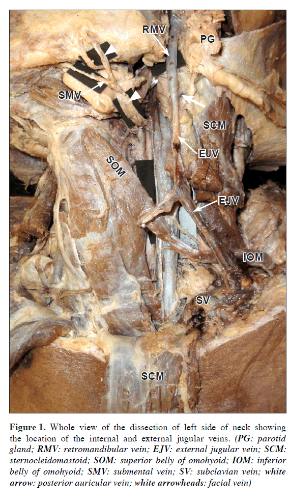
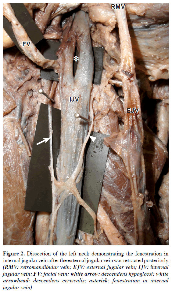
During routine dissection of head and neck region for the 1st year dental students, one female cadaver aged around 50 year showed a vertical fenestration in left IJV, the length of which was found to be 2.7 cm. The fenestration was present at the level and lateral to hyoid bone and within the carotid sheath. There were no dilatations at the level of fenestration and no structures were passing through it. In the same side near the apex of parotid gland, termination of RMV into anterior and posterior divisions was absent. Behind and inferior to the angle of mandible, the RMV joined posterior auricular vein to form the external jugular vein (EJV). The left facial vein (LFV) was running postero-inferiorly at the anterior border of masseter where it received the submental vein. Later the LFV drained directly into the left IJV at the level of its fenestration. Common facial vein was completely absent in this case. The loops of ansa cervicalis were as usual and anterior to the carotid sheath.
The venous drainage pattern of the right side of the head and neck showed no variations.
Discussion
Varying venous patterns in the region of head and neck is important for the surgeons in order to avoid any intra-operative trial and error procedures which might lead to unnecessary bleeding.
Usually the fenestrations of the arteries are associated with incidence of aneurysm at the fenestrations. This is due to turbulent flow at the bifurcation of artery or a defect in the tunica media at both the proximal and distal bifurcation points [3].
Figure 1: Whole view of the dissection of left side of neck showing the location of the internal and external jugular veins. (PG: parotid gland; RMV: retromandibular vein; EJV: external jugular vein; SCM: sternocleidomastoid; SOM: superior belly of omohyoid; IOM: inferior belly of omohyoid; SMV: submental vein; SV: subclavian vein; white arrow: posterior auricular vein; white arrowheads: facial vein)
The internal jugular vein is the continuation of sigmoid sinus. It is the largest collecting channel of head and neck. Fenestration of IJV is a rare condition and occurs in 0.4% of the population [4].
Only a very few cases of IJV fenestrations have been reported so far in the literature. The fenestrated IJV may transmit muscle or nerve or both. A case of superior belly of omohyoid and the ansa cervicalis passing through the fenestration was reported by Gonzalez-Garcia et al. [7] and spinal accessory was reported by Nayak and Soumya [8]. Fenestration of IJV has been associated with phlebectasia, a local venous dilatation [5]. In the present case no such structures were passing through IJV fenestration [2,5] and no dilatations were associated with it.
Figure 2: Dissection of the left neck demonstrating the fenestration in internal jugular vein after the external jugular vein was retracted posteriorly. (RMV: retromandibular vein; EJV: external jugular vein; IJV: internal jugular vein; FV: facial vein; white arrow: descendens hypoglossi; white arrowhead: descendens cervicalis; asterisk: fenestration in internal jugular vein)
Duplication of the IJV can be supported in the developmental aspects. The IJV developed from the precardinal vein at 3rd–6th week of intrauterine life. Any alterations in the early developmental stages of IJV may lead to its duplication [9]. According to Dodo [10], the bony partition of the jugular foramen may be due to the intrajugular process of occipital bone which passes above or behind the hypoglossal canal or due to the presence of bony partitions in the jugular foramen itself, resulting in duplication of IJV.
In the present case IJV is fenestrated in the upper part and rejoined in the lower part to form a single trunk and there is no any duplication of the IJV. Also in our case no structure was found passing through the fenestration. No clear evidence or hypothetical views for the formation of the fenestration of IJV till date have been found in the literature. A probable way for its formation may be due to the coalescence of the primitive plexus of the anterior cardinal vein into two channels during development leaving back a fenestration in its course [11].
Usually the RMV divides into its anterior and posterior divisions before emerging out at the apex of the parotid gland. The anterior division joins with the facial vein slightly inferior and anterior to the angle of mandible to form common facial vein that drains into the IJV. The posterior division unites with the posterior auricular vein to form the EJV.
In our case the RMV did not divide into its customary anterior and posterior divisions, instead it joined posterior auricular vein to form EJV. The common facial vein was absent and the facial vein drained into IJV at its fenestration. Similar cases of absence of common facial vein and facial vein draining into IJV was reported by Gonzalez-Garcia et al. [7]. Cases of facial vein draining into superficial temporal vein with an undivided RMV and termination of facial vein into the external jugular vein have been reported in literature [12].
The RMV develops from the temporal region and join the linguofacial vein to form common facial vein which drains into precardinal vein, the future IJV. EJV arises from the neck region and has connection anteriorly with facial vein and posteriorly with posterior auricular veins. In course of development, the anterior connection regresses and RMV drains into IJV via common facial vein [13]. In the present case, the RMV failed to establish the anterior connection with the facial vein to form the common facial vein, with the complete absence of anterior and posterior divisions of the RMV. The trunk of the RMV after its formation joined the posterior auricular vein to form the external jugular vein.
Knowledge of fenestration of IJV and variations in the veins of the face and neck are of immense important both clinically and surgically. Awareness of these types of variations helps to avoid radiologic misinterpretations or misidentifications of the veins of neck during the conventional radiographic procedures like angiography and catheterization or hemodialysis in renal failure patients. In cases of fenestrated IJV with the structures passing through it like, spinal accessory, superior belly of omohyoid and any other, makes it a rare variation where care should be taken during block dissections of the neck and reconstructive surgeries of brachial plexus. A sound anatomic knowledge of variations in the formation and draining patterns of the facial veins and its tributaries are essential for the microvascular surgical procedures to avoid errors leading to excessive bleeding.
References
- Hollinshead WH. Anatomy for Surgeons. Vol. 3., 3rd Ed., Jagerstown, Harper & Row. 1982; 467.
- Towbin AJ, Kanal E. A review of two cases of fenestrated internal jugular veins as seen by CT angiography. AJNR Am J Neuroradiol. 2004; 25: 1433–1434.
- Sanders WP, Sorek PA, Mehta BA. Fenestration of intracranial arteries with special attention to associated aneurysms and other anomalies. AJNR Am J Neuroradiol. 1993; 14: 675–680.
- Prades JM, Timoshenko A, Dumollard JM, Durand M, Merzougui N, Martin C. High duplication of the internal jugular vein: clinical incidence in the adult and surgical consequences, a report of three clinical cases. Surg Radiol Anat. 2002; 24: 129–132.
- Rossi A, Tortori–Donati P. Internal jugular vein phlebectasia and duplication: case report with magnetic resonance angiography features. Pediatr Radiol. 2001; 31: 134.
- Uchino A, Nomiyama K, Takase Y, Kudo S. Anterior cerebral artery variations detected by MR angiography. Neuroradiology. 2006; 48: 647–652.
- Gonzalez-Garcia R, Roman-Romero L, de la Plata MM. The rare phenomenon of internal jugular vein duplication. Otolaryngol Head Neck Surg. 2007; 137: 847-848.
- Nayak SB, Soumya KV. Neurovascular variations in the carotid triangle. International Journal of Anatomical Variations (IJAV). 2008; 1: 17–18.
- Williams PL, ed. Gray’s Anatomy. 38th Ed., Edinburgh, Churchill Livingstone. 1995; 324–327.
- Dodo Y. Observations on the bony bridging of the jugular foramen in man. J Anat. 1986; 144: 153–165.
- Setty LR. A partially double internal jugular vein and its relation to the spinal accessory nerve. J Natl Med Assoc. 1960; 52: 174–175.
- Peuker ET, Fischer G, Filler TJ. Facial vein terminating in the superficial temporal vein: a case report. J Anat. 2001; 198: 509–510.
- Mehra S, Kaul JM, Das S. Unusual venous drainage pattern of face: A case report. J Anat Soc India. 2003; 52: 64–65.
Devi Sankar K* and Sharmila Bhanu P
Department of Anatomy, Narayana Medical College, Chinthareddypalem, Nellore, Andhra Pradesh, India
- *Corresponding Author:
- Devi Sankar K, MSc Medical Anatomy
Assistant Professor, Department of Anatomy, Narayana Medical College Chinthareddy Palem, Nellore Andhra Pradesh, 524002, India
Tel: +91 949 0948006
E-mail: lesanshar@gmail.com
Date of Received: October 1st, 2010
Date of Accepted: June 14th, 2011
Published Online: August 15th, 2011
© Int J Anat Var (IJAV). 2011; 4: 144–146.
Abstract
Variations in the formation and draining patterns of the veins are common and have been frequently seen in the head and neck. A fenestration of internal jugular vein which is a rare incidence was observed in the left side neck of a cadaver during routine dissection in the Anatomy Department of Narayana Medical College. In the same side, undivided retromandibular vein and absence of common facial vein was also observed. Anatomical knowledge of such variations as found in this case is important for surgeons performing microvascular surgeries in head and neck, and also to the radiologists during their conventional radiological procedures like angioplasty, catheterization, and at times of hemodialysis in case of renal failure patients.
-Keywords
vascular fenestration, internal jugular vein, common facial vein, retromandibular vein
Introduction
Variation in the vascular system from its usual pattern is a common feature and it is more commonly observed in veins than the arteries [1]. Fenestration or incomplete divisions of cranio-cervical blood vessels are reported in the literature. Among the vessels around these regions, the arterial fenestrations are more observed than the venous fenestrations [2]. The arterial fenestrations were described in vertebral artery, basilar artery, middle cerebral artery, anterior cerebral artery, posterior inferior cerebellar artery, posterior cerebral artery, posterior communicating artery and internal carotid artery [3–6]. These fenestrated vessels do not transmit any structures through them. However, very rarely some structures may pass through these venous fenestrations.
The present article reports a case of left internal jugular vein (IJV) showing a large fenestration in the neck region, which is an uncommon finding. We also identified a variation in termination of retro-mandibular vein (RMV) and the facial vein in the same side of face and neck.
Case Report
During routine dissection of head and neck region for the 1st year dental students, one female cadaver aged around 50 year showed a vertical fenestration in left IJV, the length of which was found to be 2.7 cm. The fenestration was present at the level and lateral to hyoid bone and within the carotid sheath. There were no dilatations at the level of fenestration and no structures were passing through it. In the same side near the apex of parotid gland, termination of RMV into anterior and posterior divisions was absent. Behind and inferior to the angle of mandible, the RMV joined posterior auricular vein to form the external jugular vein (EJV). The left facial vein (LFV) was running postero-inferiorly at the anterior border of masseter where it received the submental vein. Later the LFV drained directly into the left IJV at the level of its fenestration. Common facial vein was completely absent in this case. The loops of ansa cervicalis were as usual and anterior to the carotid sheath.
The venous drainage pattern of the right side of the head and neck showed no variations.
Discussion
Varying venous patterns in the region of head and neck is important for the surgeons in order to avoid any intra-operative trial and error procedures which might lead to unnecessary bleeding.
Usually the fenestrations of the arteries are associated with incidence of aneurysm at the fenestrations. This is due to turbulent flow at the bifurcation of artery or a defect in the tunica media at both the proximal and distal bifurcation points [3].
Figure 1: Whole view of the dissection of left side of neck showing the location of the internal and external jugular veins. (PG: parotid gland; RMV: retromandibular vein; EJV: external jugular vein; SCM: sternocleidomastoid; SOM: superior belly of omohyoid; IOM: inferior belly of omohyoid; SMV: submental vein; SV: subclavian vein; white arrow: posterior auricular vein; white arrowheads: facial vein)
The internal jugular vein is the continuation of sigmoid sinus. It is the largest collecting channel of head and neck. Fenestration of IJV is a rare condition and occurs in 0.4% of the population [4].
Only a very few cases of IJV fenestrations have been reported so far in the literature. The fenestrated IJV may transmit muscle or nerve or both. A case of superior belly of omohyoid and the ansa cervicalis passing through the fenestration was reported by Gonzalez-Garcia et al. [7] and spinal accessory was reported by Nayak and Soumya [8]. Fenestration of IJV has been associated with phlebectasia, a local venous dilatation [5]. In the present case no such structures were passing through IJV fenestration [2,5] and no dilatations were associated with it.
Figure 2: Dissection of the left neck demonstrating the fenestration in internal jugular vein after the external jugular vein was retracted posteriorly. (RMV: retromandibular vein; EJV: external jugular vein; IJV: internal jugular vein; FV: facial vein; white arrow: descendens hypoglossi; white arrowhead: descendens cervicalis; asterisk: fenestration in internal jugular vein)
Duplication of the IJV can be supported in the developmental aspects. The IJV developed from the precardinal vein at 3rd–6th week of intrauterine life. Any alterations in the early developmental stages of IJV may lead to its duplication [9]. According to Dodo [10], the bony partition of the jugular foramen may be due to the intrajugular process of occipital bone which passes above or behind the hypoglossal canal or due to the presence of bony partitions in the jugular foramen itself, resulting in duplication of IJV.
In the present case IJV is fenestrated in the upper part and rejoined in the lower part to form a single trunk and there is no any duplication of the IJV. Also in our case no structure was found passing through the fenestration. No clear evidence or hypothetical views for the formation of the fenestration of IJV till date have been found in the literature. A probable way for its formation may be due to the coalescence of the primitive plexus of the anterior cardinal vein into two channels during development leaving back a fenestration in its course [11].
Usually the RMV divides into its anterior and posterior divisions before emerging out at the apex of the parotid gland. The anterior division joins with the facial vein slightly inferior and anterior to the angle of mandible to form common facial vein that drains into the IJV. The posterior division unites with the posterior auricular vein to form the EJV.
In our case the RMV did not divide into its customary anterior and posterior divisions, instead it joined posterior auricular vein to form EJV. The common facial vein was absent and the facial vein drained into IJV at its fenestration. Similar cases of absence of common facial vein and facial vein draining into IJV was reported by Gonzalez-Garcia et al. [7]. Cases of facial vein draining into superficial temporal vein with an undivided RMV and termination of facial vein into the external jugular vein have been reported in literature [12].
The RMV develops from the temporal region and join the linguofacial vein to form common facial vein which drains into precardinal vein, the future IJV. EJV arises from the neck region and has connection anteriorly with facial vein and posteriorly with posterior auricular veins. In course of development, the anterior connection regresses and RMV drains into IJV via common facial vein [13]. In the present case, the RMV failed to establish the anterior connection with the facial vein to form the common facial vein, with the complete absence of anterior and posterior divisions of the RMV. The trunk of the RMV after its formation joined the posterior auricular vein to form the external jugular vein.
Knowledge of fenestration of IJV and variations in the veins of the face and neck are of immense important both clinically and surgically. Awareness of these types of variations helps to avoid radiologic misinterpretations or misidentifications of the veins of neck during the conventional radiographic procedures like angiography and catheterization or hemodialysis in renal failure patients. In cases of fenestrated IJV with the structures passing through it like, spinal accessory, superior belly of omohyoid and any other, makes it a rare variation where care should be taken during block dissections of the neck and reconstructive surgeries of brachial plexus. A sound anatomic knowledge of variations in the formation and draining patterns of the facial veins and its tributaries are essential for the microvascular surgical procedures to avoid errors leading to excessive bleeding.
References
- Hollinshead WH. Anatomy for Surgeons. Vol. 3., 3rd Ed., Jagerstown, Harper & Row. 1982; 467.
- Towbin AJ, Kanal E. A review of two cases of fenestrated internal jugular veins as seen by CT angiography. AJNR Am J Neuroradiol. 2004; 25: 1433–1434.
- Sanders WP, Sorek PA, Mehta BA. Fenestration of intracranial arteries with special attention to associated aneurysms and other anomalies. AJNR Am J Neuroradiol. 1993; 14: 675–680.
- Prades JM, Timoshenko A, Dumollard JM, Durand M, Merzougui N, Martin C. High duplication of the internal jugular vein: clinical incidence in the adult and surgical consequences, a report of three clinical cases. Surg Radiol Anat. 2002; 24: 129–132.
- Rossi A, Tortori–Donati P. Internal jugular vein phlebectasia and duplication: case report with magnetic resonance angiography features. Pediatr Radiol. 2001; 31: 134.
- Uchino A, Nomiyama K, Takase Y, Kudo S. Anterior cerebral artery variations detected by MR angiography. Neuroradiology. 2006; 48: 647–652.
- Gonzalez-Garcia R, Roman-Romero L, de la Plata MM. The rare phenomenon of internal jugular vein duplication. Otolaryngol Head Neck Surg. 2007; 137: 847-848.
- Nayak SB, Soumya KV. Neurovascular variations in the carotid triangle. International Journal of Anatomical Variations (IJAV). 2008; 1: 17–18.
- Williams PL, ed. Gray’s Anatomy. 38th Ed., Edinburgh, Churchill Livingstone. 1995; 324–327.
- Dodo Y. Observations on the bony bridging of the jugular foramen in man. J Anat. 1986; 144: 153–165.
- Setty LR. A partially double internal jugular vein and its relation to the spinal accessory nerve. J Natl Med Assoc. 1960; 52: 174–175.
- Peuker ET, Fischer G, Filler TJ. Facial vein terminating in the superficial temporal vein: a case report. J Anat. 2001; 198: 509–510.
- Mehra S, Kaul JM, Das S. Unusual venous drainage pattern of face: A case report. J Anat Soc India. 2003; 52: 64–65.


