Variations in the Branches of Axillary Artery
Received: 09-Jun-2018 Accepted Date: Jul 04, 2018; Published: 10-Jul-2018
Citation: Ezzati M, Nasrabadi HT, Abedelahi A, et al. Variations in the branches of axillary artery. Int J Anat Var. 2018;11(3):75-76.
This open-access article is distributed under the terms of the Creative Commons Attribution Non-Commercial License (CC BY-NC) (http://creativecommons.org/licenses/by-nc/4.0/), which permits reuse, distribution and reproduction of the article, provided that the original work is properly cited and the reuse is restricted to noncommercial purposes. For commercial reuse, contact reprints@pulsus.com
Abstract
Introduction: Variations in the branching pattern of axillary artery is common. Understanding of variations of axillary artery and its branches is necessary and well documents in anatomical, radiological and surgical procedure to recognize unusual clinical signs and symptoms.
Case Report: This report reveals a case of variation in the branching pattern of axillary artery in routine dissection of female cadaver for educational purpose in Department of Anatomical sciences in Tabriz University of medical sciences.
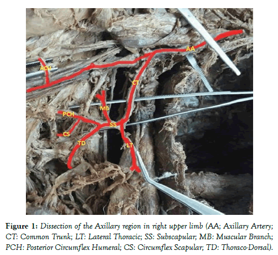
Discussion and Conclusion: Anatomically, the axillary artery divides to three parts; Superior Thoracic artery originates from the first part, lateral thoracic artery, Thoraco-acromial trunk arises from the second part, Subscapular, Anterior and Posterior Circumflex Humeral arteries arise from the third part. But in the present report we found that a common trunk including Lateral Thoracic, Subscapular arteries and muscular branches arose from the second part of axillary artery. Also in the third part we just observed Anterior Circumflex Humeral artery. A good view which clarifies the variation of axillary artery branches can prevent from the medicine mistake during radiological and surgical procedure.
Keywords
Axillary Artery; Variation; Anatomy; Dissection
Introduction
The axillary artery extends from the lateral border of the first rib as continuation of Subclavian artery then terminates at the lower border of Teres major muscle where it converses to brachial artery [1]. Most of the branches of the axillary artery supply the walls of the axillary area. Pectoralis minor muscle passes over the axillary artery and divides it to three parts. Each part also branches to few number of arteries [2]. Superior Thoracic artery originates from the first part. Lateral thoracic artery and Thoraco-acromial trunk arise from the second part. Subscapular, Anterior and Posterior Circumflex Humeral arteries arise from the third part. Subscapular artery ends into two terminal branches; Circumflex Scapular and Thoracodorsal arteries [2,3].
Case Report
Rarely variations of branching pattern in the axillary artery in right upper limb of female cadaver were found. Dissection of the axillary artery and its branches were performed according to the instructions by Cunningham’s manual of practical anatomy [4]. During a routine dissection we observed an unusual common trunk in the second part of the axillary artery. This common trunk was branched to the Lateral Thoracic, Subscapular arteries and then muscular branche. Thoracodorsal, Posterior Circumflex Humeral and Circumflex Scapular arteries arose from Subscapular artery. The final branch of the axillary artery was Anterior Circumflex Humeral artery, which was the only branch of the third part (Figure 1).
Discussion and Conclusion
Shanta Kumar et al. reported about the variation in branching of axillary artery approximately similar to our finding. Actually they found that the second part of the right axillary artery gave rise to a common trunk in 50 years old male cadaver. So that Lateral thoracic and subscapular arteries originated from this common trunk. On the other hand, anterior and posterior circumflex humeral arteries arose from the third part of the axillary. Interestingly the left axillary artery had normal condition without any variation [5]. In the other case aastha et al. found that the first and third part of the axillary artery had variation in an adult male cadaver. According to their reports the first part gave superior thoracic artery then it divided to superior and inferior branches. The superior one supplied first intercostal space and the inferior branch moved to the second intercostal space. Also the third part gave a common trunk with several branches including Subscapular, anterior and posterior circumflex humeral arteries. The reasons of the variation in normal branching pattern of axillary artery may be due to the defects of surrounding tissue and also defects in embryonic vascular network development [6]. In the other study the axillary artery in 25 cadavers was exposed. They investigated the variation of its branches and then reported that one of the specimens had different branching which result in the axillary artery was branched superficial and deep trunk. In this rare observation all branches originated from deep trunk including superior thoracic, lateral thoracic, thoraco-acromial arteries. In addition, deep part of artery divided into anterior and posterior branches in which the continuation of anterior one branched to anterior circumflex humeral, posterior circumflex humeral and profunda brachii arteries. Then the posterior part became subscapular artery and divided into two branches of circumflex scapular and thoraco-dorsal arteries. The remarkable point is that ulna and radial arteries related to the branches of superficial trunk [7]. Kanaka et al. studied the axillary artery in 30 cadavers. They reported the branching pattern as: 1) posterior circumflex humeral artery from subscapular artery, 2) subscapular artery and thoraco-acromial trunk along with each other from the second part of axillary artery, and 3) accessory subscapular artery from the third part of axillary artery (duplex origin) were common variation which observed in those cadavers [8]. Heulke investigated 89 cadavers, then reported his data and compared it with other author’s works. In this case, he showed that subscapular artery was usually branch of third (79.2%) or the second (15.7%) part of the axillary artery. But it was rarely reported that the subscapular artery originated from the first part of the axillary artery (0.6%) or from brachial artery (2.8%), which is the one of axillary divisions. In addition the circumflex scapular and thoracodorsal arteries, the subscapular artery could be the common trunk for the lateral thoracic artery (14.1%) and posterior circumflex humeral artery (15.2%) [9]. All of these documents about anatomical variations can prevent from the medicine mistake during radiological and surgical procedure and also promote treatment method in clinical situation.
Acknowledgement
Authors of this paper wish to appreciate who donate their body to promote the education and research. Written informed consent was obtained from Department of Anatomical sciences. Hamid Tayefi Nasrabadi accepts full responsibility for this work.
REFERENCES
- Snell RS. Clinical anatomy: Lippincott Williams & Wilkins. 2004.
- Standring S. Gray’s Anatomy E-Book: The Anatomical Basis of Clinical Practice: Elsevier Health Sciences. 2015.
- Tank PW, Grant JCB. Grant’s dissector: Lippincott Williams & Wilkins. 2012.
- Cunningham D. Cunningham’s Manual of Practical Anatomy volume 1 Upper and Lower Limbs. 1976.
- Shantakumar SR, Mohandas Rao K. Variant branching pattern of axillary artery: a case report. Case reports in vascular medicine. 2012.
- Aastha AJ, Kumar MS. An Unusual Variation of Axillary Artery: A Case Report. JCDR. 2015;9:AD05.
- Sawant SP, Shaikh ST, More RM. The Study of variations in the branches of axillary artery. Inter J Adv Physiol Allied Sci. 2012;1:1-7.
- Kanaka S, Eluru RT, Basha MA, et al. Frequency of Variations in Axillary Artery Branches and its Surgical Importance. IJSS. 2015;3:1-4.
- Huelke DF. Variation in the origins of the branches of the axillary artery. The Anatomical Record. 1959;135:33-41.


