Variations of circle of Willis in human cadavers
Received: 06-Mar-2018 Accepted Date: Mar 22, 2018; Published: 03-Apr-2018, DOI: 10.37532/1308-4038.18.11.43
Citation: Shubhangi Y. Variations of circle of Willis in human cadavers. Int J Anat Var. 2018;11(2):43-45.
This open-access article is distributed under the terms of the Creative Commons Attribution Non-Commercial License (CC BY-NC) (http://creativecommons.org/licenses/by-nc/4.0/), which permits reuse, distribution and reproduction of the article, provided that the original work is properly cited and the reuse is restricted to noncommercial purposes. For commercial reuse, contact reprints@pulsus.com
Abstract
Aims and Objective: The blood supply of brain is of great importance because of the metabolic demands of the nervous tissue. A significant anastomosis, the Circle of Willis exists between the carotid and vertebral arterial systems. An understanding of the distribution of the arteries is very important. As the neurological signs depends on the site of lesion.
Material and Methods: The study was conducted on 45 adult brain specimens of both sexes in human cadavers. The collected specimens were preserved in 10% formalin. The Circle of Willis of each brain was dissected with care.
Results: Variations of Circle of Willis in brain specimens includes – anomalous morphology, incomplete circle, and variable shape of Circle and variable length and external diameter of arteries of Circle of Willis. In the present study the terminal bifurcation of the basilar artery was found to be unequal in few cases.
Conclusion: Various diseases of arteries of brain like cerebrovascular attack, aneurysm, haemorrhage etc. are related to the anatomic patterns of the Circle of Willis. The knowledge of which is of considerable help to neurosurgeons.
Keywords
ACA; PCA; PCoA; Basilar artery
Introduction
The brain gets a copious arterial supply from a pair of internal carotid and a pair of vertebral arteries. Both arterial systems form a polygonal anastomosis, the Circle of Willis, at the base of the brain around the interpenduncular fossa. The internal carotid arteries supply the frontal, parietal and part of the temporal lobes, and the vertebral arteries through the basilar artery and its terminal branches supply the occipital and part of temporal lobes, together with the brain stem and the cerebellum [1].
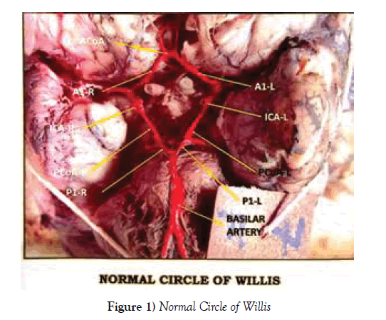
The arterial circle is formed by the internal carotid artery which is interconnected by the anterior cerebral arteries (ACA) on both the sides and an anterior communicating artery (ACoA) which connects the right and the left anterior cerebral arteries. The carotid system is connected to the posterior cerebral arteries (PCA) of the vertebral system by two posterior communicating arteries (PCoA).
The Circle of Willis equalizes the blood flow to the different parts of the brain and under normal condition little interchange of blood takes place across the anastomotic channel due to equality of the blood pressure. The streams of blood conveyed by the carotid and vertebral systems meet in the posterior communicating artery at a ‘dead point’ where the pressure of the two is equal and no admixture of blood occurs. However, in case of occlusion of one of the arterial systems, the blood crosses the middle line through the communicating branches and maintains nutrition of the opposite brain by contralateral flow. Therefore, the Circle of Willis acts as principal collateral channel to preserve the independent cerebral blood flow when normal, or dependent blood flow in occlusion of one of the main arterial feeders.
As the neurological signs depend on the site of lesion, an understanding of the distribution of the arteries is very essential. A detailed knowledge on various configuration of the Circle of Willis is also important for surgical interventions.
Arterial occlusion by an embolus or thrombus is usually followed by infarction of a region supplied. Aneurysms often develop at the sites of branching of arteries which can rupture or leak causing subarachnoid haemorrhage.
Usually the occipital lobe is supplied by the posterior cerebral artery (PCA). If precommunicating part of PCA is larger than the posterior communicating artery (PCoA), then the occipital lobe is supplied by vertebra – basilar artery. But sometimes the precommunicating part of PCA may be smaller than PCoA, then occipital lobe is mainly supplied by internal carotid artery (Standring 2008) [1].
Inspite of immense advances in technology through computerized tomographical angiography or magnetic angiography, the dissection based study has its own importance for the study of anatomy.
The aim of the study was to evaluate the anatomic configuration of the Circle of Willis in north Indian population and to compare the results with previously conducted research.
Functional demands of the growing brain tissue govern the development of cerebral vasculature. With the regression of some earlier vessels and appearance of new channels, a fully developed circle becomes functional at the end of 6-7 weeks. As the arterial circle reaches its typical configuration, the occurrence of multiple events during the embryological stage might lead to a different spectrum of vascular anatomical variants [2]. Duplications and fenestrations of an artery can take place due to an incomplete fusion of arteries [3].
Materials and Methods
The present study was conducted on 45 adult brain specimens of human cadavers. The specimens were collected from cadavers given for dissection in dissection hall of Department of Anatomy and autopsies done in the department of forensic medicine. The collected specimens were preserved in 10% formalin. The Circle of Willis of each brain was dissected out with care. The detailed study of segments of the arterial circle was done in each specimen and the findings were noted with reference to:
1) Shape of the arterial circle.
2) Position and course of all the arteries forming the Circle of Willis.
3) Measurements: Vernier calipers, graduated to measure up to 0.1 mm, were used to measure the length and the external diameter of the components of the Circle of Willis.
The measurements of the external diameter of the following vessels were taken:
a) Anterior communicating artery (ACoA): at the midpoint;
b) Right and left anterior cerebral artery (A1): close to their origin;
c) Right and left internal carotid artery (ICA): between the origin of anterior cerebral and posterior communicating artery.
d) Right and left posterior communicating arteries (PCoA): at the mid-point;
e) Right and left posterior cerebral arteries (P1): at the midpoint from its origin and the anastomotic termination of PCoA.
f) Basilar artery (BA): close to its end.
The arteries measuring less than 1 mm in diameter were considered to be abnormal whereas the communicating arteries less than 0.5 mm in diameter were considered to be abnormal. The measurement of the lengths of the components of the Circle of Willis was taken:
a) Right and left A1 segment from the point of its origin to the point of the ACoA communication;
b) Anterior communicating artery from the point of communication between right A1 segments to the left A1 segment;
c) Right and left PCoA from the point of their origin to the point of termination.
d) Right and left P1 segment from their origin at the bifurcation of the basilar artery to the point of communication of the PCoA.
The arterial circle was colored with premium gloss enamel red color paint by using a ‘0’ number brush. Photographs were taken.
Results and Observations
Completeness of the circle of Willis
In the present study, the Circle of Willis was found to be complete in 37 (82.22%) cases. Out of 37 complete circles 13 were symmetric and 24 asymmetric circles.
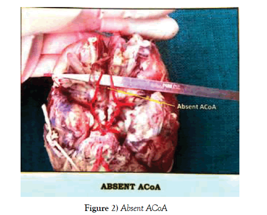
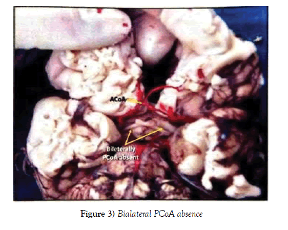
It was observed that the circle was incomplete in 8 of the 45 brain specimens (17.78%). Out of which the anterior part of the circle was found to be incomplete in 2 cases (4.44%) while the posterior part was incomplete in 6 cases (13.33%).
Shape of the circle of Willis
In the present study, the shape of the circle in most was a nonagon found in 33 cases (73.33%) and in 5 cases the circle was a polygon (11.11%).
In the present study the anterior part of the circle was anomalous in 7 specimens (15.56%); while the posterior part was more anomalous than anterior part which was seen in 24 cases (53.33%).
External diameter and length of the components
In the present study the external diameter and length of the components of the Circle of Willis was measured. The average of the external diameter and the length of the components are as follows: (Tables 1 and 2).
| Name of the arterial segment | Length (mm) | Diameter (mm) | ||
|---|---|---|---|---|
| Right | Left | Right | Left | |
| BA | _ | 3.6 ± 0.12 | ||
| P1 | 7.98 ± 0.42 | 8.02 ± 0.45 | 2.04 ± 0.07 | 2.18 ± 0.14 |
| PCoA | 13.98 ± 0.44 | 14.46 ± 0.49 | 1.14 ± 0.09 | 1.21 ± 0.08 |
| ICA | _ | 3.65 ± 0.24 | ||
| A1 | 13.99 ± 0.44 | 14.40 ± 0.12 | 2.02 ± 0.04 | 2.08 ± 0.05 |
| ACoA | 3.98 ± 0.34 | 2.98 ± 0.12 | ||
Table 1: Average length and external diameter in the present study
| Basilar artery | No. of specimens | Percentage | |
|---|---|---|---|
| Bifurcation | Equal | 39 | 86.66% |
| Unequal | 6 | 13.33% | |
| BA ⇨ PCA | Right | 3 | 6.66% |
| Left | 8 | 17.77% | |
Table 2: Basilar Artery
In the present study, the terminal bifurcation of the basilar artery was found to be equal in 39 cases (86.66%); unequal in 6 cases (13.33%). Out of the 6 unequal cases it was found that the basilar artery continued as left PCA in 2 and right PCA in 4 cases (Figures 1-3).
Discussion
The cerebral arterial circle and its branches are subjected to numerous variations. The arteries forming the Circle of Willis also vary in size. The findings were compared with the previous workers on this subject as follows:
1. Circle Morphology: Completeness
Kapoor K et al. [4] examined the circle of willis in 1000 medico legal autopsies. Out of 1000 specimens examined, 452 (45.2%) conformed to the typical pattern. In the rest of the specimens (54.8%) there were variations. The circle was deficient in 32 cases (3.2%).
Bergman et al. represented that the normal Circle of Willis which is a complete polygon show considerable variability not only in its components but also of its branches [5].
Wieslawa K et al. measured the diameters of cerebral vessels by a slide caliper in 100 brain specimens of polish population. 27% of cases presented the typical pattern. The remaining 73% of all cases were atypical; in 16% the circles was incomplete and in 57% complete [6].
2. Circle Morphology: Shape
Fisher et al. observed that the prevalence of the typical normal arterial polygon ranges from 4.6% to 72.2% [7].
According to Guerin et al., Hillen, the shape of the Circle of Willis is determined at the time of development of the supplying vessels during the first two decades of life [8,9].
3. Circle Morphology: Anomalous
PN Jain et al., conducted a study on 144 formalin fixed human brains and found that the total number of specimens showing anomalies were 115 (80.55%). Anterior and posterior parts of the Circle of Willis showed anomalies in 42 (29.16%) and 74 (51.38%) cases respectively. These findings correlate with the findings of the present study [10].
4. Basilar Artery (BA)
Kamath S (1979) in a study on 100 Circle of Willis observed the average external diameter of basilar artery to be 3.5 mm. He also found that abnormally narrow diameter was found most frequently in posterior cerebral artery followed by posterior communicating arteries [11].
Padmavati G (2006) in a study on Circle of Willis found the average external diameter of basilar artery to be 4.8 mm [12].
So from the above discussion, it is clear that the Circle of Willis have lot of morphological variations.
Conclusion
In the present study, all the components of the Circle of Willis were dissected and studied in detail and the variations were noted down. All the available literature of the studies performed on Circle of Willis previously was reviewed and the findings were compared with the present study. The fact that the Circle of Willis has lot of morphological variations is accepted universally. There is lot more left to be studied on this topic. The genetical factors having role in the variations of the Circle are not known clearly.
The various diseases of carotid and vertebral arterial systems like CVA (stroke), hemorrhage, aneurysm etc. are related to the anatomic patterns of the Circle of Willis. So the study of normal as well as variations is very important for us to understand and find the location of the lesion, the knowledge of which is of great importance for the neurosurgeons.
REFERENCES
- Susan Standring. The Anatomical basis of clinical practice. Gray’s Anatomy. 2008;39:296-301.
- Milenkovic Z, Vucetic R, Puzic M. Assymmetry and Anomalies of the Circle of Willis in fetal brain, microsurgical study and functional remarks. Surgical Neurol. 1985;24:563-70.
- Menshawi K, Mohr JP, Gutierrez J. A Functional Perspective on the Embryology and Anatomy of the Cerebral Blood Supply. J Stroke. 2015;17:144-58.
- Kapoor K, Singh B, Dewan LI. Variations in the configuration of the circle of Willis. Anat Sci Int. 2008;83: 96-106.
- Bergman RA, Afifi AK, Miyaucchi R. Compendium of human anatomic variations text atlas and world literature. Baltimore Urban and Schwarenberg. 1988;pp:66-8.
- Wieslawa Klimek P, Monika R, Aleksandra W, et al. A multitude of variations in the configuration of the circle of Willis: an autopsy study. Anat Sci Int Sep. 2016;91:4:325-33.
- Fisher CM. The circle of Willis: Anatomical variations. Vasc Dis. 1965;2:99-105.
- Guerin J, Gouaze A, Lazorthes G. Le polygone de Willis de l’enfant et les facteurs de son modelage. Neurochirurgie. 1976;22:217-26.
- Hillen B. The Variability of the Circulus Arteriosus (Willisii): order or anarchy. Acta Ant. 1987;129:74-80.
- Jain PN, Kumar V, Thomas RJ, et al. Anoamalies of human cerebral arterial circle (of Willis). J Anat Soc of India. 1990;39:137.
- Kamath S. A study of the dimensions of the basilar artery in South Indian subjects. J Anat Society of India.1979;28: 45-64.
- Padmavatee G. Study of Basilar Artery in Human Cadevers. 2006.


