Variations of hepatic circulation in two human cadavers with clinical implications
2 Rush College of Health Sciences, Rush University, Chicago IL, USA
3 Department of Cell and Molecular Medicine, Rush University, Chicago IL, USA, Email: christopher_ferrigno@rush.edu
Received: 30-Aug-2018 Accepted Date: Sep 20, 2018; Published: 27-Sep-2018
Citation: Katrikh AZ, Maheia T, Ferrigno C. Variations of hepatic circulation in two human cadavers with clinical implications. Int J Anat Var. Sep 2018;11(3):109-110.
This open-access article is distributed under the terms of the Creative Commons Attribution Non-Commercial License (CC BY-NC) (http://creativecommons.org/licenses/by-nc/4.0/), which permits reuse, distribution and reproduction of the article, provided that the original work is properly cited and the reuse is restricted to noncommercial purposes. For commercial reuse, contact reprints@pulsus.com
Summary
Two rare variations of hepatic vasculature were discovered during routine anatomical dissection in the gross anatomy laboratory at Rush University. The first case is that of an 82-year-old female with the left hepatic artery as a branch of the left gastric artery. The right lobe of the liver was supplied by the right hepatic, in turn from the common hepatic artery, as normal. The second case is that of a 95-year-old male with an accessory right hepatic artery that arose directly from the celiac trunk. The accessory right hepatic artery coursed posterior to the portal triad, giving off the cystic artery on its way to the right lobe of the liver. The cystic artery was located in Calot’s triangle as normal, however it coursed posterior in the sagittal plane. Knowledge of variations in vasculature to the liver is of great importance to hepatobiliary surgeons and both diagnostic and interventional radiologists.
Keywords
Right hepatic artery; Left hepatic artery; Accessory right hepatic artery
Traditionally, the liver receives vasculature from the right hepatic artery (RHA) and left hepatic artery (LHA), which are indirect branches originating from the celiac trunk. In this foregut-derived paradigm, the celiac trunk gives off three principle arteries including the hepatic artery, splenic artery, and left gastric artery. The common hepatic artery further bifurcates into two branches, the gastroduodenal which projects inferiorly and the proper hepatic artery which projects superolaterally towards the liver and associated structures. It is the proper hepatic artery which, after the right gastric artery branches off, ultimately gives off the LHA and RHA, with the right hepatic artery serving the gallbladder via its branch, the cystic artery. A meta-analysis conducted in 2004 of two seminal works and other subsequent studies of hepatic artery distribution suggests that traditional vascular supply is present in 52%–79.1% of cases [1-3]. This study further suggests that the most prevalent variant of hepatic artery distribution occurs when the RHA originates from the superior mesenteric artery, a finding in 3.5%–18% of subjects. In 2.5%–14.2% of cases, the LHA branches off the left gastric artery. The authors of the meta-analysis subsequently conducted their own examination of 604 abdominal angiographies and described 15 cases of an LHA off of the left gastric and only a single case of an accessory right hepatic artery originating from the celiac trunk. In rare cases, an LHA off of the left gastric artery wraps around the esophagus which causes a challenge for surgical ligation during liver transplant [4]. Additionally, a study done in 2016 showed that an aberrant LHA was present in 15% of laparoscopic Nissen fundoplication cases [5]. Variations were reported in a 2017 cadaveric case report that described an individual with a celiac trunk giving off an accessory RHA in addition to the left gastric artery, hepatosplenic trunk (common origin of common hepatic and splenic arteries), and left inferior phrenic artery [6]. A cadaveric study also published in 2017 described 3 individuals with, in addition to a primary RHA stemming from the proper hepatic artery, an accessory RHA as the fourth branch of the celiac trunk. The same study went on to describe 2 more individuals where the accessory RHA was a branch of the celiac trunk, which also gave off the left gastric artery and a hepatosplenic trunk [7]. More recently, a retrospective analysis done in 2018 of abdominal CT contrast angiography showed a new anatomical variation of a celiac trunk with 7 branches, one of which was a RHA that did not arise from the proper hepatic [8]. Additionally, a laparoscopic cholecystectomy case report in 2016 described an individual with a cystic artery which stemmed directly from the celiac trunk that did not course through Calot’s triangle [9]. Calot’s triangle is an anatomical space composed of the cystic duct, common hepatic duct, and inferior border of the liver. The triangle is used as a landmark during cholecystectomy procedures to locate and ligate the cystic artery, which is traditionally located within that space.
Case Report
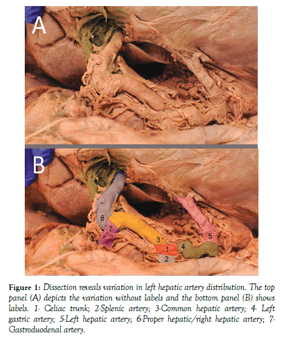
Two rare variations of hepatic vasculature were discovered during routine anatomical dissection in the instructional gross anatomy laboratory at Rush University. In both instances, the donors expired due to causes unrelated to the hepatic circulation. The first case is that of an 82-year-old female with the LHA branching off the left gastric artery (Figure 1). The RHA/proper hepatic branched off the common hepatic artery as normal. Past surgical history was positive for a cholecystectomy; therefore, the cystic artery had been ligated and removed prior to dissection.
Figure 1: Dissection reveals variation in left hepatic artery distribution. The top panel (A) depicts the variation without labels and the bottom panel (B) shows labels. 1- Celiac trunk; 2-Splenic artery; 3-Common hepatic artery; 4- Left gastric artery; 5-Left hepatic artery; 6-Proper hepatic/right hepatic artery; 7- Gastroduodenal artery.
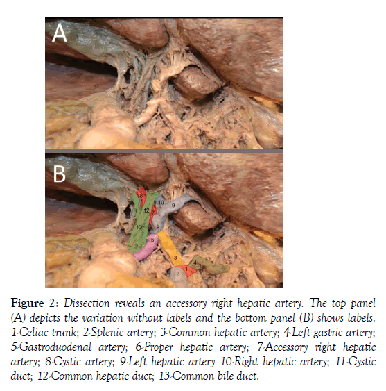
The second case is that of a 95-year-old male that had an accessory RHA that arose directly from the celiac trunk (Figure 2). The accessory RHA coursed posterior to the portal triad, giving off the cystic artery on its way to the right lobe of the liver. The cystic artery was located in Calot’s triangle as normal, however its course was posterior to the triangle, in the sagittal plane.
Figure 2: Dissection reveals an accessory right hepatic artery. The top panel (A) depicts the variation without labels and the bottom panel (B) shows labels. 1-Celiac trunk; 2-Splenic artery; 3-Common hepatic artery; 4-Left gastric artery; 5-Gastroduodenal artery; 6-Proper hepatic artery; 7-Accessory right hepatic artery; 8-Cystic artery; 9-Left hepatic artery 10-Right hepatic artery; 11-Cystic duct; 12-Common hepatic duct; 13-Common bile duct.
Discussion
The anatomical variation described in the 82-year-old female is that of the LHA as a branch of the left gastric artery. This variation has been described in literature, and although rare, is prevalent enough to warrant classification as a type II variation by the works of Michels and Hiatt [2,3]. This variant can affect gastrectomy and esophagectomy procedures, where part or all of the left gastric artery distribution is ligated. The aberrant LHA in this case would have to be spared and anastomosed to remaining vasculature so that blow could flow to the left hepatic lobe. Additionally, the artery is located in a position susceptible to injury during hiatal hernia repair. Great care must be taken to preserve the LHA to prevent ischemia to the hepatic left lobe, especially if the artery is not the accessory, but rather the primary vascular supply.
The anatomical variation described in the 95-year-old male is significantly rarer than the first. An accessory RHA as a branch of the celiac trunk was not described by the original works of Michels and Hiatt [2,3]. In our review of the literature, we were only able to find eight instances of an accessory RHA off the celiac trunk ever described (1,6-9). Of these, only four cases have the same variation [1,7]. None of these studies described whether the cystic artery was a branch of the normal or accessory RHA. Although our cystic artery was a branch of the accessory and coursed through Calot’s triangle, a similar aberration gave some difficulty to a cholecystectomy procedure in 2016 [9], and therefore this variation merits further description. An RHA that does not arise directly from the proper hepatic artery may cause an aberrant coursing pattern en route to the gall bladder, which can have clinical consequences, particularly during laparoscopic cholecystectomy which has become one of the most common general surgery procedures.
Knowledge of the types and rates of vascular variations to the liver is of great importance to any surgeon working in the abdomen, especially for hepatobiliary procedures. Additionally, this information is relevant to both diagnostic and interventional radiologists.
Acknowledgement
We would like to thank the individuals who graciously and generously donate their remains to Anatomical Gift Association of Illinois for education and research purposes. We would like to thank James Williams, PhD and Anthony Serici, the Director and Lab Manager, respectively, of the Rush University Anatomy laboratory. We would also like to thank Jeffery Nelson, MD for assistance in preparing the figures.
REFERENCES
- Koops A, Wojciechowski B, Broering DC, et al. Anatomic variations of the hepatic arteries in 604 selective celiac and superior mesenteric angiographies. Surg Radiol Anat. 2004;26:239-44.
- Michels NA. Newer anatomy of the liver and its variant blood supply and collateral circulation. Am J Surg. 1996;112:337-47.
- Hiatt JR, Gabbay J, Busuttil RW. Surgical anatomy of the hepatic arteries in 1000 cases. Ann Surg. 1994;220:50-2.
- Altaca G, Karakayali H, Haberal M. An extremely uncommon variation of left hepatic artery around the esophagus: A case report. Clin transplant. 2009;23:121-3.
- Hendrickson RJ, Yu S, Bensard DD, et al. Preservation of an aberrant left hepatic artery during laporascopic nissen fundoplication. J Soc Laparoend. 2016;10:180-3.
- Daescu E, Sztika D, Lapadatu AA, et al. Rare variant of celiac trunk branching pattern associated with modifications of hepatic arterial vascularization. Rom J Morphol Embryol. 2017;58:969-75.
- Olewnik L, Wysiadecki G, Polguj M, et al. Types of coeliac trunk branching including accessory hepatic arteries: A new point of view based on cadaveric study. Folia Morphol. 2017;76:660-7.
- Rusu MC, Manta BA. Novel anatomical variation: Heptafurcation of the celiac trunk. Surg Radiol Anat. 2018;40:457-63.
- Katagiri H, Sakamoto T, Okumura K, et al. Aberrant right hepatic artery arising from the celiac trunk: A potential pitfall during laparoscopic cholecystectomy. Asian J Endosc Surg. 2016;9:72-4.


