Ellis-Van Creveld Syndrome with partial atrioventricular canal defect: A case report and review of literature
Received: 19-Jan-2018 Accepted Date: Jan 24, 2018; Published: 15-Feb-2018
Citation: Nagre SW, Abhilash, Ravikumar V, et al. Ellis-Van Creveld Syndrome with partial atrioventricular canal defect: A case report and review of literature. Gen Surg: Open Access. 2018;1(1):1-3.
This open-access article is distributed under the terms of the Creative Commons Attribution Non-Commercial License (CC BY-NC) (http://creativecommons.org/licenses/by-nc/4.0/), which permits reuse, distribution and reproduction of the article, provided that the original work is properly cited and the reuse is restricted to noncommercial purposes. For commercial reuse, contact reprints@pulsus.com
Abstract
Ellis Van Creveld Syndrome [EVC] is a rare genetic disorder also as called chondroectodermal dysplasia. It has autosomal recessive inheritance caused by mutation in the EVC gene located on the short arm of chromosome 4. It mainly consists of four components-Chondro dysplasia in the form of dwarfism, ectodermal dysplasia mainly in teeth and nail, polydactyly and congenital heart disease. It is commonly seen in the the Amish population of Pennsylvania in USA but also occur in non Amish population with the prevalence around 7/1,000,000 live births. Here we report a case of 15 year old Indian female born to a consanguineous marriage with the classical features of Ellis Van Creveld Syndrome diagnosed with a partial AV canal defect having a relatively asymptomatic childhood . A Partial AV canal defect is an uncommon cardiac malformation, and yet it is commonly found in patients with the EVC. We operated on her with septal patching and anterior mitral leaflet cleft repair with smooth recovery.
Introduction
Richard W.B. Ellis of Edinburgh and Simon van Creveld of Amsterdam met in a train compartment while traveling to a pediatrics conference in England in the late 1930s and discovered that each had a patient with the specific similar pathology because of chondroectodermal dysplasia and named it Ellis van Creveld Syndrome. It is an autosomal recessive syndrome with disproportionate dwarfism, postaxial polydactyly, ectodermal dysplasia, a small chest, and a high frequency of congenital heart defects . Other features include partial harelip and multiple frenulae in the lips; short ribs and narrow chest; genital abnormalities like epispadias, hypospadias and cryptorchidism; low iliac wings with spur-like projections at acetabula and genu valgum [1]. Although most patients have normal intelligence, occasional central nervous system anomalies or mental retardation has been reported. It is commonly seen in the Amish population of Pennsylvania in USA but also occur in non Amish population with the prevalence around 7/1,000,000 live births [2]. Here we report a case of 15 year old Indian female born to a consanguineous marriage with the classical features of Ellis Van Creveld Syndrome diagnosed of a partial AV canal defect with a cleft in AML and having a relatively asymptomatic childhood. A Partial AV canal defect is an uncommon cardiac malformation, and yet it is commonly found in patients with the EVC. We operated on her with septal patching and anterior mitra leaflet cleft repair on CPB with smooth recovery.
Case Report
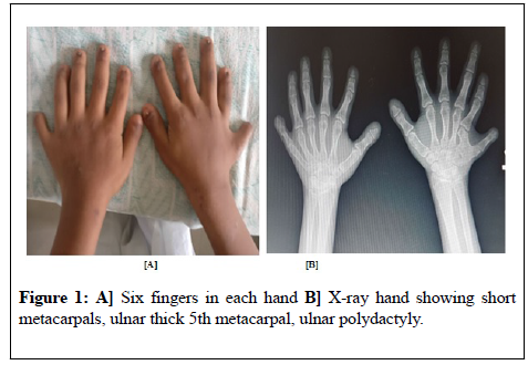
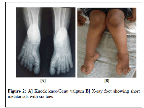
A 15 years young female presented to us with progressive exertional dyspnea and easy fatigability of six months duration with no history of orthopnea or PND. She had no significant illness in the past except that she was born with short limbs, six fingers in each hand and six digits in each foot [Figure 1A]. She also has a history of frequent respiratory tract infections. She had short stature which was apparent from two years of age. Her school performance was below average. She attained menarche early at the age of ten years and having normal menstrual cycles once in a month since then with no h/o any other bleeding tendencies. On examination, she was found to have a short stature with a height of 115 cm. Her younger sister also, was having short stature. The finger and toe nails were small and brittle. Oral cavity examination revealed absent incisors and the rest were natal teeth. The patient had knock knees [Figure 2A] with pectus excavatum. Examination of cardio-respiratory system revealed narrow chest and had a prominent diastolic thrill at the apex, a loud S1 with a low-pitched grade 4/6 mid-diastolic rumbling murmur and a pansystolic murmur in the mitral area. The S2 was widely split, fixed with a loud P2. There was a grade 3/6 ejection systolic murmur over the left second intercostal space. Other systems were normal. All blood investigations are normal. Skeletal radiology of upper limb revealed short metacarpals, ulnar thick 5th metacarpal, ulnar polydactyly [Figure 1B] and X-ray foot showed short metatarsals with six toes [Figure 2B].
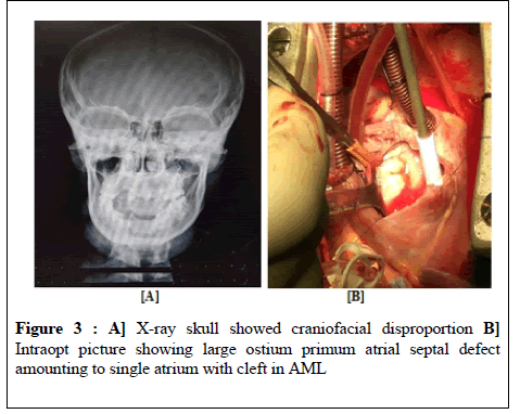
Radiological examination of the lower limb revealed depressed and flattened lateral compartment of upper end of tibia on both sides giving rise to genu valgum. X-ray skull showed craniofacial disproportion [Figure 3A]. Chest X-ray showed cardiomegaly. Ultrasound abdomen was normal. Electrocardiogram showed normal sinus rhythm, first degree heart block with biatrial enlargement. Echocardiography showed partial AVCD-large ostium primum atrial septal defect amounting to single atrium, two separate atrioventricular valves with moderate mitral regurgitation, cleft in AML,right atrium and right ventricle dilated, mild pulmonary artery hypertension. Catheterization study showed classical Goose Neck deformity due to elongated left ventricular outflow tract in the left ventricular angiogram. The saturation run showed significant step up in the right atrium with QP/QS-5.65. Mean pulmonary artery pressure was recorded as 30 mm Hg. We operated on her with pericardial patch closure of ostium primum atrial septal defect and repair of cleft in AML with 5-0 prolene through right atriotomy on cardiopulmonary bypass after arresting the heart under GA [Figure 3B]. Postopt recovery was uneventful. Echo was done s/o patch insitu with no residual shunt and no mitral regurgitation. The patient was discharged on the seventh postopt day after stitch removal with advice regular advice of six monthly followup. Recent follow up showed that the patient’s symptoms have reduced with increased tolerance for work.
Discussion
Six-fingered dwarfism ('digital integer deficiency') was an alternative designation used for this condition when it was being studied in the Amish and may have served a useful function in defining this then little known condition for the medical profession, as well as the lay public. The term, however, has been found offensive by some, apparently not because of 'dwarfism,' but because of the reference to the polydactyly, which is seen as a 'freakish' labelling [3]. For this reason, 6-fingered dwarfism has been removed as an alternative name for this entry. This leaves Ellis–van Creveld syndrome with its initialism, EVC, as the only satisfactory designation. It is caused by mutation in the EVC gene located on chromosome 4 short arm [4]. Emerging molecular and developmental studies suggest that EVC and EVC2 proteins may be important for cilia function, which is implicated in the pathogenesis of heterotaxy syndromes. It is speculated that coordinate function between the EVC proteins is required for a cilia-dependent cardiac morphogenesis [5]. The parents are the carriers of the mutation and there is a 25% chance of further pregnancies resulting in a child with a similar problem.
Nagre et al. has discussed the ostium primum ASD in detail in his studies [6,7]. Cardiac manifestation are seen in 50-60% of cases. Common atrium (CA) is an uncommon cardiac malformation, and yet it is commonly found in patients with EVC [8]. Patient had a complete atrioventricular canal defect which is the most common congenital abnormality seen in 40% of cases followed by partial atrioventricular canal defect, a ventricular septal defect or a patent ductus arteriosus [9]. Liver and Central nervous system abnormalities could also occur rarely in Ellis Van Creveld syndrome. Other genitourinary anomalies like renal agenesis, renal tubular dilatation, nephrocalcinosis, megaureters, vulvar atresia. 50% patients with Ellis Van Creveld syndrome usually die in infancy due to recurrent respiratory infections. Those without any cardiac abnormality may have a normal life expectancy. The patient having a complete atrioventricular canal defect with EVC has survived till 19 years without any significant pulmonary artery hypertension [10]. There are only few reports of cardiac surgical repairs done for this condition. Our patient was born to a consanguineous marriage with similar EVC syndromic features in her younger sister. After diagnosis of PAVCD we investigated the patient in a multidisciplinary way and then operated as a routine cardiac surgery with no intraopt or postopt complications.
Conclusion
Ellis van Creveld syndrome, though rare in Indian population, but with a multidisciplinary approach with involvement of dentist, psychologist, paediatrician, urologist, orthopedician, pulmonologist, cardiologist, cardiac surgeon and occupational therapist management seems to be easy and simple. Early diagnosis and treatment of cardiac abnormality in EVC syndrome patients will definitely decrease the morbidity and mortality with improved quality of life.
Ethical approval: All procedures performed in studies involving human participants were in accordance with the ethical standards of the institutional and/or national research committee and with the 1964 Helsinki declaration and its later amendments or comparable ethical standards.
REFERENCES
- Phatak SV, Kolwadkar PK, Phatak MS. Ellis van Creveld syndrome: Report of two cases. Indian J Radiol Imaging 2003;13:393-4.
- Natal teeth. Medline Plus : U.S. National Library of Medicine 2013.
- Ellis-van Creveld syndrome. Medline Plus : U.S. National Library of Medicine 2013.
- Ikramullah K, Syed A, Kiren M. Ellis van C reveld syndrome: A case report. J Pak Assoc Derma 2006;16:239-242.
- Ruiz-Perez VL, Goodship JA. Ellis–van Creveld syndrome and Weyers acrodental dysostosis are caused by cilia-mediated diminished response to hedgehog ligands. Am J Med Genet C Semin Med Genet 2009;151:341-351.
- Nagre SW. Atrial septal defect-management approach in children - Annals of woman and child health, Letter to Editor 2006;1-2.
- Nagre SW, Nagre MS. Observational study of surgical closure of ostium primum atrial septal defect in thirty paediatric patients.Annals of woman and child health 2015;1:1-4.
- Baujat G, Le Merrer M. Ellis van C reveld syndrome: Review. O rphanet Journal of Rare Dis 2007;2:27: 1-5.
- McKusick VA, Egeland JA, Eldridge R, Krusen DE. Dwarfism in the Amish I. The Ellis–van Creveld syndrome. Bulletin of the Johns Hopkins Hospital 1964;115: 306-36.
- Toshifumi M, Takehiro K, Jun-ichi O et al. Left atrioventricular valve regurgitation after repair of incomplete atrioventricular septal defect' Ann Thoac Surg 2004;77: 2157-2162.


