The role and correlation analysis of PI3K/AKT pathway in breast carcinoma with lymph node metastasis
2 Department of Breast Surgery, The Affiliated Hospital of Taishan Medical University, Tai’an 271000, Shandong Province, China, Email: dangxiangguo@yahhoo.com
Received: 10-Oct-2018 Accepted Date: Oct 24, 2018; Published: 30-Oct-2018
Citation: Wang Y, Dang X, Zhang M, et al. The role and correlation analysis of PI3K/AKT pathway in breast carcinoma with lymph node metastasis. J Immunopathol 2018;1(1): 4-7.
This open-access article is distributed under the terms of the Creative Commons Attribution Non-Commercial License (CC BY-NC) (http://creativecommons.org/licenses/by-nc/4.0/), which permits reuse, distribution and reproduction of the article, provided that the original work is properly cited and the reuse is restricted to noncommercial purposes. For commercial reuse, contact reprints@pulsus.com
Abstract
Discuss the correlation of the expressions of PI3K, AKT2 and VEGFR-3 protein in breast carcinoma and the role of lymph node metastasis.
Methods: Of the 236 breast carcinoma cases in total were selected for immunohistochemistry. The expressions of PI3K, AKT2 and VEGFR-3 protein were detected by methods immunohistochemical SP. The expressions of PI3K, AKT2 and VEGFR-3 mRNA were detected by RT-PCR.
Results: The positive rates of PI3K in patients with lymph node metastasis was 71.09% (91/128), which was significantly higher than that in patients without lymph node metastasis group (58.33%, 63/108, χ2=6.6802, P=0.0097) ). AKT2 and VEGFR-3 expressions were also significantly higher in lymph node metastasis group than that of the non-lymph node metastasis group, 71.09% ( 91/128) vs.52.78%, (57/108) (χ2=9.3474, P=0.0022) and 92.97% (119/128) vs. 83.33% (90/108) (χ2=5.3676, P=0.0205. Of the 236breast cancer patients, the relative expression levels of PI3K, AKT2 and VEGFR-3 mRNA in invasive ductal breast carcinoma with lymph node metastasis were0.62 ± 0.21, 0.49 ± 0.18 and 0.55 ± 0.22 respectively, significantly higher than that without lymph node metastasis0.49 ± 0.15 and 0.59 ± 0.22 and 0.49 ± 0.15,the difference between two groups was statistically significant(P=0.0475, 0.0007 and 0.0364). The linear correlation analysis showed that the relative expressions of PI3K mRNA and VEGFR-3 mRNA in breast cancer were positively correlated (r=0.5, P <0.001). Similarly, the relative expressions of AKT2 mRNA and VEGFR-3 mRNA were also positively correlated (r=0.46, P <0.001).
Conclusion: The relative expressions of PI3K mRNA and VEGFR-3 mRNA in breast cancer were positively correlated. The up-regulation of PI3K and AKT2 may promote the over-expression of VEGFR-3 in breast cancer cells, induce lymphangiogenesis and promote lymphatic metastasis.
Introduction
Extensive dissemination of primary breast cancer is the leading cause of death, and axillary lymph nodes is the first step in extensive metastasis, lymph node metastasis has become one of the main criteria for the prognosis of breast cancer patients and treatment options [1]. Clinical experiments show that tumor cells migrating to lymph nodes must pass lymphatic vessels and new lymphangiogenesis process and greatly promote the lymphatic metastasis of tumor. The key protein that induces lymphangiogenesis is vascular endothelial growth factor receptor 3 (VEGFR-3), which is activated by vascular endothelial growth factors C and D (VEGF-C and VEGF-D), creating a new favorable environment for lymphangiogenesis [2]. Phosphatidylinositol 3-kinase/serine protein kinase B (PI3K/AKT) signaling pathway is dysregulated in most human tumors and regulates tumor cell proliferation and apoptosis and is involved in IGF-1 induction VEGF-C up-regulation is an important role in the process of lymphatic metastasis of breast cancer [3]. AKT2 is one of the isoform of AKT, as an oncogene, AKT2 protein is overexpressed in many tumor tissues [4]. However, the mechanism of the interaction between PI3K/AKT expression in breast cancer and lymphatic metastasis is not completely understood. In this dissertation, we examined the relationship between PI3K/AKT protein and VEGFR-3 expression in breast cancer by detecting the expression of PI3K/AKT, to investigate the role of its expression on lymphatic metastasis of breast cancer.
Materials and Methods
Specimen
General information: The project has been approved by the Medical Ethics Committee of the Affiliated Hospital of Taishan Medical College. Between 1/1/2015 and 12/31/2017, 236 female patients with pathologically proven invasive ductal carcinoma from the Department of Breast Surgery, Affiliated Hospital of Taishan Medical College. By HE staining pathological diagnosis of invasive ductal carcinoma of breast. It including 128 patients with lymph node metastasis and 108 patients without lymph node metastasis. The patients’ median age was 47 (43.6 ± 3.1) years old (range 27-66 years old). All patients had no prior radiotherapy or chemotherapy. Based on tumor TNM staging from the International Union Against Cancer (UICC) and American Cancer Society (American Joint Committee on Cancer, AJCC), 38 cases were stage Ⅰ, 113 cases were stage Ⅱ, and 85 cases were stage Ⅲ. Detailed pathological features are shown in Table 1.
| lymph node metastasis | n | PI3K | AKT2 | VEGFR-3 | ||||||
|---|---|---|---|---|---|---|---|---|---|---|
| Positive | χ2 | P | Positive | χ2 | P | Positive | χ2 | P | ||
| lymph node metastasis | 128 | 95(74.22) | 6.6802 | 0.0097 | 91(71.09) | 9.3474 | 0.0022 | 121(92.97) | 5.3676 | 0.0205 |
| without lymph node metastasis | 108 | 63(58.33) | 57(52.78) | 86(79.63) | ||||||
Table 1: Expressions of pi3k, akt2and vegfr-3 protein in breast invasive ductal carcinoma NNT(%)
For the experiment purpose, approximately 1.0 cm × 1.0 cm × 1.0 cm fresh specimen from each case was taken and stored at -80oC refrigerator. The remaining specimens were fixed with formaldehyde and embedded in paraffin for pathological diagnosis.
Main reagents: Rabbit anti-human PI3K (PI3K p85) polyclonal antibody, rabbit anti-human AKT (AKT2 isoform)polyclonal antibody, rabbit antihuman VEGFR-3 polyclonal antibody were bought in Signalway Antibody, trypsin, streptavidin- horseradish peroxidase (SP) kit (sp-9001) and concentrated DAB color kit were purchased from Beijing Golden Bridge Biotechnology Co., Ltd., one-step RT-PCR kit was purchased from Beijing Tiangen Biochemical Technology Co., Ltd.. Chemiluminescence reagents were purchased from Santa Cruz. The pre-stained protein marker was purchased from Solarbio. Trizol reagents were purchased from Invitrogen, and the RT-PCR primers were synthesized by Invitrogen (Shanghai).
PI3K and AKT2 and VEGFR-3 protein detection
Specimens from 236 patients were embedded in paraffin and sectioned 4μm thick. Immunohistochemical SP method was used to remove the endogenous peroxidase. Antigens were retrieved by high temperature and high pressure and non-specific binding was blocked with goat serum. The slides were then incubated with primary antibodies at room temperature, followed by a biotin-labeled secondary antibody. Tissues were then stained with 3,3′-diaminobenzidine (DAB). Finally, tissues were counterstained with hematoxylin, dehydrated, and mounted. The slides were reviewed under the light microscope within 48 hours. We used PI3K and AKT2-positive sections provided by the reagent company as positive controls. For negative controls, we incubated slide with PBS instead of primary antibodies.
Interpretation of staining results
PI3K and AKT2 located in the cytoplasm were stained as yellow to brown granules, while VEGFR-3 positive cells showed brown particles in cytoplasm or on cell membrane. For each group, known positive tissue was used as positive control and PBS was used instead of one antibody as negative control. Two pathologists evaluated the slides by double-blind method. Three fields from each slide were randomly selected and the positive cells per 100 tumor cells per field were recorded at X400. The final result from each slide was the average of the three fields. Grading standards were based on reference [5] with modification: (1) Staining intensity scores: no stain: 0 point, light yellow: 1 point, brown yellow: 2 points, brown: 3 points; (2) Percentage of positive cells: Positive cells <25% were counted as 0 point; 26~ 50% is 1 point;51% ~ 75% is two points;>75% is 3 points, and the final criterion is the score value of the product of the two points. Immunohistochemical results: 0 ~ 1 is divided into the negative (-), and 2 ~ 3 into the positive (+~ + +), more than 4 points for strong positive (+ + +).
PI3K and AKT2 and VEGFR-3 mRNA expression
Total RNA was extracted from fresh specimens (236 cases) by TRIzol method. The total RNA was extracted from each tissue. Total RNA (2μg) from each case was reversely transcribed using Reverse transcription polymerase chain reaction (RT-PCR) and bet-2microglobulin (B2M) was used as internal references. The gene of PI3K, AKT2, VEGFR-3 and internal reference gene B2M were designed and synthesized by Shanghai Invitrogen Bioengineering Co., Ltd. by using the software of Primer5.0. The primer used as follows: B2M primer F: 5’-GGCTATCCAGCGTACTCCAAA-3 ‘, R: 5’-CGGCAGGCATACTCATCTTTT-3 ‘, product length 377bp; VEGFR-3 primer sequence F: 5’-TGTGGAGGGAAAGAATAAGAC-3’, R: 5’-CCTCTAGTAGCTCCTCGGAT-3 ‘, product length 182bp; PI3K primer sequence F: 5’-CATCAC TTC CTC CTG CTC TAT-3 ‘, R: 5’-CAGTTGTTGGCAATCTTCTTC-3’, product length 246 bp; AKT2 primer sequence F: 5’-GCGACGTGGCTATTG TGA-3 ‘, R: 5’-CAGTCTGGATGGCGGTTG-3’, product length 545 bp. The optimal PCR condition for VEGFR-3 and B2M was 94°C for 4 min followed by 32 cycles of 94°C for 30s, 60°C for 30s, 72°C for 1 min and 72°C for additional 7 min. The optimal PCR condition for PI3K and AKT2 was 94°C for 4 min followed by 30 cycles of 94°C for 30 s, 58°C for 30 s and 72°C for 1 min and 72°C for additional 7 min. Amplification products were separated on a 2.0% agarose gel electrophoresis and photographed under a UV transmission instrument. Negative result was defined by no electrophoretic band at the position corresponding to the molecular weight of marker, and positive result was defined by presence of target band at the corresponding position of marker molecular weight. The image analysis software BandScan version 4.3 was used to analyze the optical density of the electrophoresis band. The ratio of the Optical density value of each target band to the internal reference B2M was the relative light expression of the target genes PI3K, AKT2 and VEGFR-3.
Statistical methods
SPSS 17.0 statistical analysis software, the experimental data χ2 test and linear (level) correlation analysis. Measurement data results to ± S said, between groups were compared using one-way analysis of variance and t test, the test level a=0.05.
Results
PI3K and AKT2 and VEGFR-3 expression in breast cancer tissue
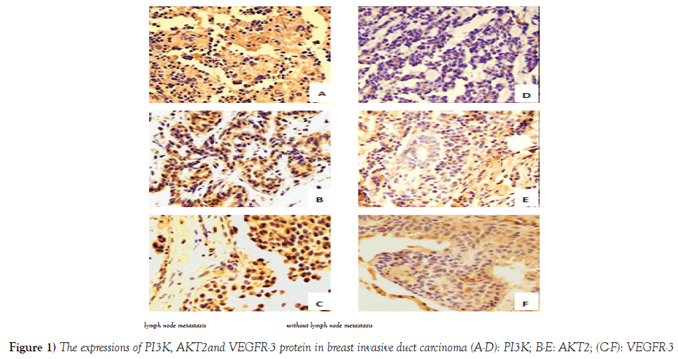
Of the 236 breast cancer cases, positive rates of PI3K, AKT2 and VEGFR-3 were 66.95% (158/236), 61.86% (146/236) and 88.56% (209/236), respectively. The positive rate of PI3K in patients with lymph node metastasis was 71.09% (91/128), which was significantly higher than that in patients without lymph node metastasis group (58.33%, 63/108, χ2=6.6802, P=0.0097) ). AKT2 and VEGFR expressions were also significantly higher in lymph node metastasis group than that of the non-lymph node metastasis group, 71.09% ( 91/128) vs.52.78%, (57/108) (χ2=9.3474, P=0.0022) and 92.97% (119/128) vs. 83.33% (90/108) (χ2=5.3676, P=0.0205, Figure 1 and Table 1), respectively.
PI3K and AKT2 and VEGFR-3 mRNA expression and correlation
Of the 236 breast cancer patients, the relative expression levels of PI3K, AKT2 and VEGFR-3 mRNA in invasive ductal breast carcinoma with lymph node metastasis were significantly higher than that without lymph node metastasis (P=0.0475, 0.0007 and 0.0364).
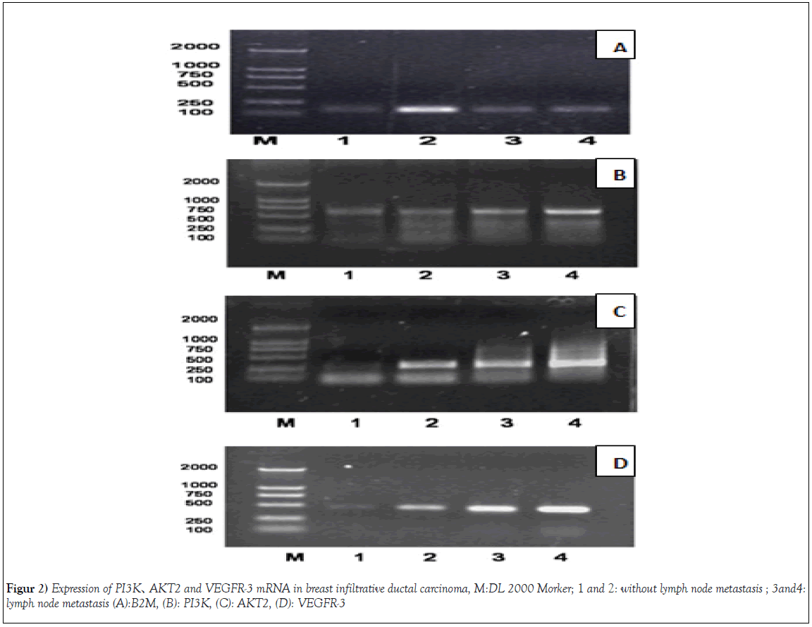
The linear correlation analysis showed that the relative expressions of PI3K mRNA and VEGFR-3 mRNA in breast cancer were positively correlated (r=0.5, P <0.001). Similarly, the relative expressions of AKT2 mRNA and VEGFR-3 mRNA were also positively correlated (r=0.46, P <0.001, Table 2 and Figure 2).
| lymph node metastasis | n | PI3KmRNA | P | AKT2mRNA | P | VEGFR-3mRNA P | |
|---|---|---|---|---|---|---|---|
| lymph node metastasis | 128 | 0.62 ± 0.21 | 0.0475 | 0.49 ± 0.18 | 0.0007 | 0.55 ± 0.22 | 0.0364 |
| without lymph node metastasis | 108 | 0.49 ± 0.15 | 0.59 ± 0.22 | 0.49 ± 0.15 | |||
Table 2: The expressions of PI3K、AKT2 and VEGFR-3mRNA protein in breast carcinoma.
Discussion
Breast cancer is one of the most common malignant tumors in women from Europe and the United States. According to the latest statistics, China has a relatively low incidence of breast cancer as compare to the United States and Canada, with 2.16 breast cancer per million females Cancer, while the mortality rate reached 15.74% [6]. Lymphatic metastasis of breast cancer is based on lymphangiogenesis. VEGFR-3 is a key protein that induces lymphangiogenesis and a specific marker of lymphatic endothelial cells. VEGFR-3 is an important factor of VEGF-C, D / VEGFR-3 signaling pathway. Experiments confirmed that VEGFR-3 is a receptor tyrosine kinase, VEGFR-3 overexpression and VEGF-C and VEGF-D binding, through the signal transduction, induced lymphatic endothelial cell proliferation, migration and lymphangiogenesis, promoting breast cancer cell survival and metastasis [7]. The spread of the tumor to the lymph nodes and lymphatic system is an important pathway. The expression of VEGF-C induces lung lymphangiogenesis and promotes the lung metastasis of breast cancer. This pattern of metastasis suggests that the VEGF-C / VEGFR-3 pathway not only serves as a preventive measure to the goal of metastasis but also the treatment to established metastatic disease [8].
PI3K / AKT signaling pathway in most human cancers dysregulation, not only can regulate tumor cell proliferation and apoptosis, but also tumor angiogenesis, lymphangiogenesis and invasion and metastasis. All kinds of breast cancer subtypes have different degrees of PI3K / AKT signaling pathway activation and the activation of the pathway indicates a poor prognosis. PI3K is activated in breast cancer tissue which an important component of breast cancer signal transduction pathway [9]. Among the two signals, AKT2 is an important cross between multiple signaling pathways. Under physiological conditions, AKT2 exists in the cytoplasm and stays low activity. Under the stimulation of various factors, activated AKT2 and phosphorylation (p-AKT2) plays a role in affecting multiple downstream effector molecules as well as in the occurrence, development, infiltration and metastasis of breast cancer. Especially in estrogen receptor-positive breast cancer, AKT2 activation is very common, which has a clear correlation with histological grade and lymph node metastasis, suggesting that the high expression of AKT2 may play a role in the malignant transformation and metastasis of breast cancer Important role, also indicates the endocrine therapy resistance [10]. However, AKT2 morphology functions specially in different stages of breast cancer progression. AKT1 participates in local tumor growth and AKT2 participates in distant tumor transmission. AKT2 has a low prognostic value and is therefore a valuable target for therapy [11]. With the exception of involvement in cell transformation, tumorigenesis, progression of cancer, and resistance to breast cancer; mutations in the PI3K/ AKT pathway are evident in breast cancer [12]. The relationship between PI3K/AKT expression with clinicopathological features and the prognosis of breast cancer patients was found to be significantly correlatedwith axillary lymph node metastasis [13]. In invasive ductal carcinoma of the breast, AKT2 also up-regulates the expression of VEGF-C, thereby promoting lymph node metastasis in breast cancer. It is speculated that PI3K/AKT/VEGF-C pathway may play a role in lymphatic metastasis of invasive ductal carcinoma of breast [14].
Of the 236 breast cancer cases, the expression of PI3K, AKT2 and VEGFR-3 in breast cancer tissues with lymph node metastasis was significantly higher than that in patients without lymph node metastasis group (P<0.05 or P<0.01 ). RT-PCR also showed that the relative expression of PI3K, AKT2 and VEGFR-3 mRNA in invasive ductal breast cancer with lymph node metastasis were significantly higher than that without lymph node metastasis (P<0.05 or P<0.01). Moreover, the relative expressions of PI3K mRNA 、AKT2 mRNA and VEGFR-3 mRNA in breast cancer were positively correlated (P<0.001). It is speculated that the up-regulation of PI3K and AKT2 in breast cancer may promote the over-expression of VEGFR-3 in breast cancer cells and induce the lymphatic metastasis through the PI3K/AKT pathway system in lymphatic endothelial cells and induce lymphangiogenesis.
The occurrence and development of breast cancer are not the result of a single gene effect, nor are they involved in the PI3K/AKT pathway. The future treatment of breast cancer should also be a combination of inhibition or activation of multiple gene pathways in order to achieve the desired effect [15]. We suggest that the PI3K/AKT and VEGF-C and D/VEGFR-3 pathways play a common role in the lymphatic metastasis of invasive ductal carcinoma of the breast and regulate the pathways and lymphangiogenesis, thereby promoting lymphatic endothelial cell growth and lymph node metastasis occur in breast cancer. As a result, it will turn hopes into reality that the PI3K/AKT signal pathway and related proteins as targets for therapy of breast cancer, in order to improve the therapeutic effect of breast cancer, inhibit the lymphatic metastasis of breast cancer, to improve the patient’s survival.
Acknowledgements
This work was supported by the National Natural Science Foundation of China (No.81473687), and Natural Science Foundation of Shandong Province, China (No.ZR2009CM039 and No. ZR2013HM038), and Health Science and Technology Subject of Shandong Province (No. 2016BJ0015), and Science and Technology plan of Tai’an (No.2015NS2082).
REFERENCES
- Yonemura Y, Endou Y, Tabachi K, et al. Evaluation of lymphatic invasion in primary gastric cancer by a new monoclonal antibody,D2-40. Hum Pathol. 2006;37(9):1193-9.
- Ran S, Volk L, Hall K, et al. Lymphangiogenesis and lymphatic metastasis in breast cancer. Pathophysiology. 2010;17(4):229-51.
- Zhu C, Qi X, Chen Y, et al. PI3K/AKT and MAPK/ERK1/2 signaling pathways are involved in IGF-1-induced VEGF-C upregulation in breast cancer. J Cancer Res ClinOncol. 2011;137(11):1587-94.
- Edge, Stephen Byrd B, David Compton R, et al. AJCC Cancer Staging Manual,7th ed[M]. New York: Springer-Verlag New York Inc, 2010;347-76.
- Chau NM, Ashcroft M. AKT: a role in breast cancer metastasis. Breast Cancer Res. 2004;6(1):55-7.
- Fan L, Strasser-Weippl K, Li JJ, et al. Breast cancer in China. Lancet Oncol. 2014;15(7):e279-e89.
- Kurenova EV, Hunt DL, He D, et al. Vascular endothelial growth factor receptor-3 promotes breast cancer cell proliferation, motility and survival in vitro and tumor formation in vivo. Cell Cycle. 2009;8(14):2266-80.
- Das S, Ladell DS, Podgrabinska S, et al. Vascular endothelial growth factor-C induces lymphangitic carcinomatosis, an extremely aggressive form of lung metastases. Cancer Res,2010;70(5):1814-24.
- Lopez KE Mcneil CM, Millar EK, et al. PI3K pathway activation inbreast cancer is associated with the basal-like phenotype and cancerspecific mortality. Int J Cancer. 2010;126(5):1121-31.
- Ma CX, Suman V, Goetz MP, et al. A Phase II Trial of Neoadjuvant MK-2206, an AKT2 Inhibitor, with Anastrozole in Clinical Stage II or III PIK3CA-Mutant ER-Positive and HER2-Negative Breast Cancer. Clin Cancer Res. 2017;23(22):6823-32.
- Riggio M, Perrone MC, Polo ML, et al. AKT1 and AKT2 isoforms play distinct roles during breast cancer progression through the regulation of specific downstream proteins. Sci Rep. 2017;7:44244.
- Guerrero-Zotano A, Mayer IA, Arteaga CL. PI3K/AKT/mTOR: role in breast cancer progression, drug resistance, and treatment. Cancer Metastasis Rev. 2016;35(4):515-524.
- Wang LL, Hao S, Zhang S, et al. PTEN/PI3K/AKT protein expression is related to clinicopathological features and prognosis in breast cancer with axillary lymph node metastases. Hum Pathol 2017;61:49-57.
- Liang N, Li Y, Chung HY. Two natural eudesmane-type sesquiterpenes from Laggera alata inhibit angiogenesis and suppress breast cancer cell migration through VEGF- and Angiopoietin 2-mediated signaling pathways. Int J Oncol. 2017;51(1):213-22.
- Choi SK, Kim HS, Jin T. Overexpression of the miR-141/200c cluster promotes the migratory and invasive ability of triple-negative breast cancer cells through the activation of the FAK and PI3K/AKT signaling pathways by secreting VEGF-A. BMC Cancer. 2016;2(16):570.
Keywords
Breast carcinoma; lymphatic metastasis; lymphangiogenesis; pi3k/akt pathway; vascular endothelial growth factor receptor-3(VEGFR-3)


