Unilateral duplex ureter
Received: 14-Mar-2018 Accepted Date: Apr 03, 2018; Published: 16-Apr-2018, DOI: 10.37532/1308-4038.18.11.51
Citation: Tang MJ, Arachchi A, Mehdipour R, et al. Unilateral duplex ureter. Int J Anat Var. 2018;11(2):51-52.
This open-access article is distributed under the terms of the Creative Commons Attribution Non-Commercial License (CC BY-NC) (http://creativecommons.org/licenses/by-nc/4.0/), which permits reuse, distribution and reproduction of the article, provided that the original work is properly cited and the reuse is restricted to noncommercial purposes. For commercial reuse, contact reprints@pulsus.com
Abstract
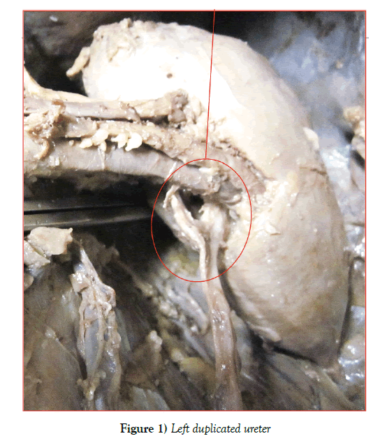
A routine cadaveric dissection of a 77 year old female with metastatic breast cancer demonstrated an ectopic unilateral left duplex ureter. The duplicated ureters originated separately at the renal pelvis, communicating 2 cm below the originating point. This anatomical variant is estimated to represent 1% of all renal tract anomalies, occurring in 0.5% of the total population. The clinical outcomes of duplex ureters are well documented in current literature.
Keywords
Cadaveric dissection; Ureters; Kidney; Gonadal vessels
Introduction
We describe a case of left unilateral ectopic duplex ureter, which was noted during a routine dissection of a cadaver. Multiple complications of this anatomical variation have been previously reported.
The ureter is the connecting vessel between the kidney and bladder, measuring 25-30 cm in length. It is composed of smooth muscle lined internally with a mucosal membrane [1,2]. It begins proximally at the renal pelvis, posterior to the renal artery and vein. With regards to the left ureter, this usually corresponds to the level of the second lumbar vertebra while the right is marginally lower. The ureter descends along the medial surface of Psoas major, passing retroperitoneal and above the genitofemoral nerve before being crossed by the gonadal vessels [2]. The proximal section of the right ureter lies posterior to the duodenum while the inferior section is crossed by the right colic, ileocolic and superior mesenteric vessels. The left ureter continues lateral to the inferior mesenteric vessels, entering the pelvis anterior to the sacroiliac joint, at the bifurcation of the common iliac vessels. Here it adheres to the posterior abdominal peritoneum. In females, the ureter is then traversed by the Infundibulo pelvic ligament anteriorly, before travelling medially and entering the base of the bladder [2].
The ureter is not uniform in diameter. Its calibre is narrowest at three points: the pelvo- ureteric junction, the section overlying the pelvic brim and its termination at the bladder. Renal calculi frequently become lodged at these anatomical landmarks [3].
Embryologically, the ureteric bud forms from the mesonephric duct. Duplication of the renal pelvis (Partial duplication) arises when the bud diverges above the vesico- ureteric junction before reaching the metanephric blastema [4]. In complete ureteral duplication, the development of two ureteral buds from a single mesonephric duct gives rise to two separate ureters and renal pelves [5]. It is theorised that duplication is environmentally influenced or genetically driven, determined by incomplete penetrance of an autosomal dominant trait [6,7].
Case Report
As depicted in our image, there is an ectopic duplex ureter arising from the left renal pelvis which communicates with the original ureter as they descend below the renal vessels. The aberrant ureter is smaller in diameter compared with the original and measures 2 cm from origination to intersection. There is only one additional ureter noted and no ectopic ureter was identified on the right side. Routine cadaveric dissection revealed no further anatomical abnormality (Figure 1).
Discussion
The duplex ureter is a relatively common anatomical abnormality, constituting 1% of all renal tract malformations [8]. They are classified as complete or incomplete, depending on whether a communication exists between the two tubes. The majority of incomplete duplex ureters originate separately at the renal pelvis, as in our case, before communicating and entering the bladder as one entity. This variant is rarely associated with clinical abnormality [4]. Duplication may also occur at the middle and lower third of the ureter, entering the bladder at two separate insertion points [4]. When the convergence point occurs at the lower third, peristalsis of urine freely occurs between both ureters, resulting in compromised transmission of urine to the bladder. This in turn, leads to dilation of the upper urinary system, urinary stasis, loin pain and increased risk of developing infection and renal calculi [1]. Interestingly, Wakhlu and colleagues determined that renal function was not impacted by the site of ectopic ureter insertion [9].
Complete duplex ureters do not communicate at all, with separate originating and insertion points at the renal pelvis and bladder respectively. It is not known if the incidence and severity of urological complications are different in complete and incomplete duplex ureters
Duplex ureters are usually detected on prenatal ultrasound. Those which are not, may present with loin pain, ureterocele, vesicouretic reflux and recurrent urinary tract infection at an early age [10,11]. Late onset development of UTI is rare. Additionally, this anatomical variant is a well recognised cause of urinary incontinence, particularly in females, though urinary incontinence in a boy with duplex ureters has previously been reported by Ejaz and colleagues [1,12].
It is possible for patients to remain asymptomatic for many years or indefinitely. The former often present with renal calculi obstruction difficult to identify on regular investigation. Nyanhongo et al. described a case of a 39 year old male with a 6mm non-obstructing calculus identified on noncontrast CT KUB. Consequent ureteroscopy failed to isolate the calculus as it was situated in the previously unidentified ectopic ureter [8].
Conclusion
Duplex ureters are a relatively common congenital malformation with varying clinical consequence. During surgical management and clinical assessment, it is important to be aware of these variations especially when first line investigation fails to identify a cause for the presenting symptoms. This variant has previously been reported in existing literature and its clinical significance has been well documented. Awareness of this variation may aid in diagnosis of unexplained urological symptoms when traditional treatment fails to improve the presenting complaint.
Acknowledgement
The authors would like to thank all who contributed to this paper including the University of Melbourne Anatomical department for providing cadavers for dissection.
REFERENCES
- Duplex ureter. Lancet. 1988;5:515.
- Last RJ, RMH McMinn. Last's Anatomy, Regional and Applied.
- Ordon M, Schuler TD, Ghiculete D, et al. Stones lodge at three sites of anatomic narrowing in the ureter: clinical fact or fiction?. Endourol. 2013;27:270-6.
- Kogan BA. Disorders of the ureter and ureteropelvic junction. Gen Urol. 2008.
- Karakose A, Aydogdu O, Atesci YZ. Unilateral complete ureteral duplication with distal ureteral stone: A rare entity. Can Urol Assoc J. 2013;7:511-2.
- Atwell JD, Cook PL, Howell CJ, et al. Familial incidence of bifid and double ureters. Arch Dis Child. 1974;49: 825-6.
- Phillips DIW, Divall JM, Maskell RM, et al. A geographical focus of duplex ureter. Br J Urol. 1987;60:329-31.
- Nyanhongo DK, Antil, S, Nasir, S. Pitfalls of the duplex system: the mystery of the missing stone. BMJ Case Reports. 2017.
- Wakhlu A, Dalela D, Tandon RK, et al. The single ectopic ureter. Br J Urol. 1998;82:246-51.
- Van den Hoek J, Montagne GJ, Newling DW. Bilateral intravesical duplex system ureteroceles with multiple calculi in an adult patient. Scand J Urol Nephrol. 1995; 29:223-4.
- Raja J, Mohareb AM, Bilori B. Recurrent urinary tract infections in an adult with a duplicated renal collecting system. Radiol Case Rep. 2016;11:328-31.
- Ejaz T, Malone PS. Male duplex urinary incontinence. J Urol. 1995;153:470-1.


